Lifestyle change, stress management can reduce Alzheimer’s risk

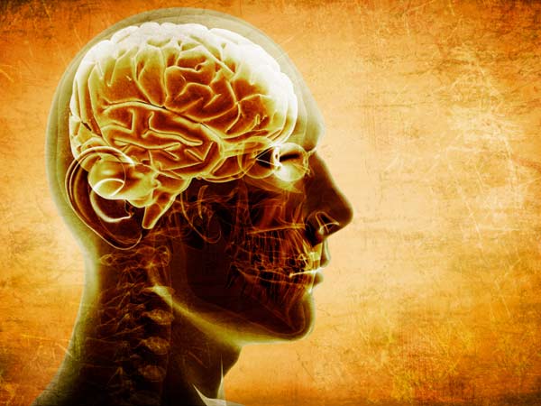
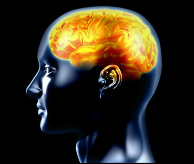
Change in lifestyle accompanied by proper stress management and indulgence in cognitive activities and socialising can help manage and reduce the risk factors for Alzheimer’s disease, experts said.

Lifestyle change, stress management can reduce Alzheimer's risk

Though Alzheimer’s is not curable, but lifestyle modifications like engaging in physical activity, eating the right diet and socialising can delay the onset of the disease, they said.

“Alzheimer’s though is not curable as yet, being involved in activities that keep the brain active is the best way to mitigate the risks associated with Alzheimer’s,” said Manjari Tripathi, professor of neurology at AIIMS.

Tripathi was speaking to reporters at a conference on “How to reduce risks of Alzheimer’s and the new innovations in its treatment”, organised by the Delhi chapter of the Alzheimer’s and Related Disorders Society of India (ARDSI). ARDSI in collaboration with the Netherlands embassy will Sep 19 host a discussion on Alzheimer’s and also release the World Alzheimer’s Report 2014.

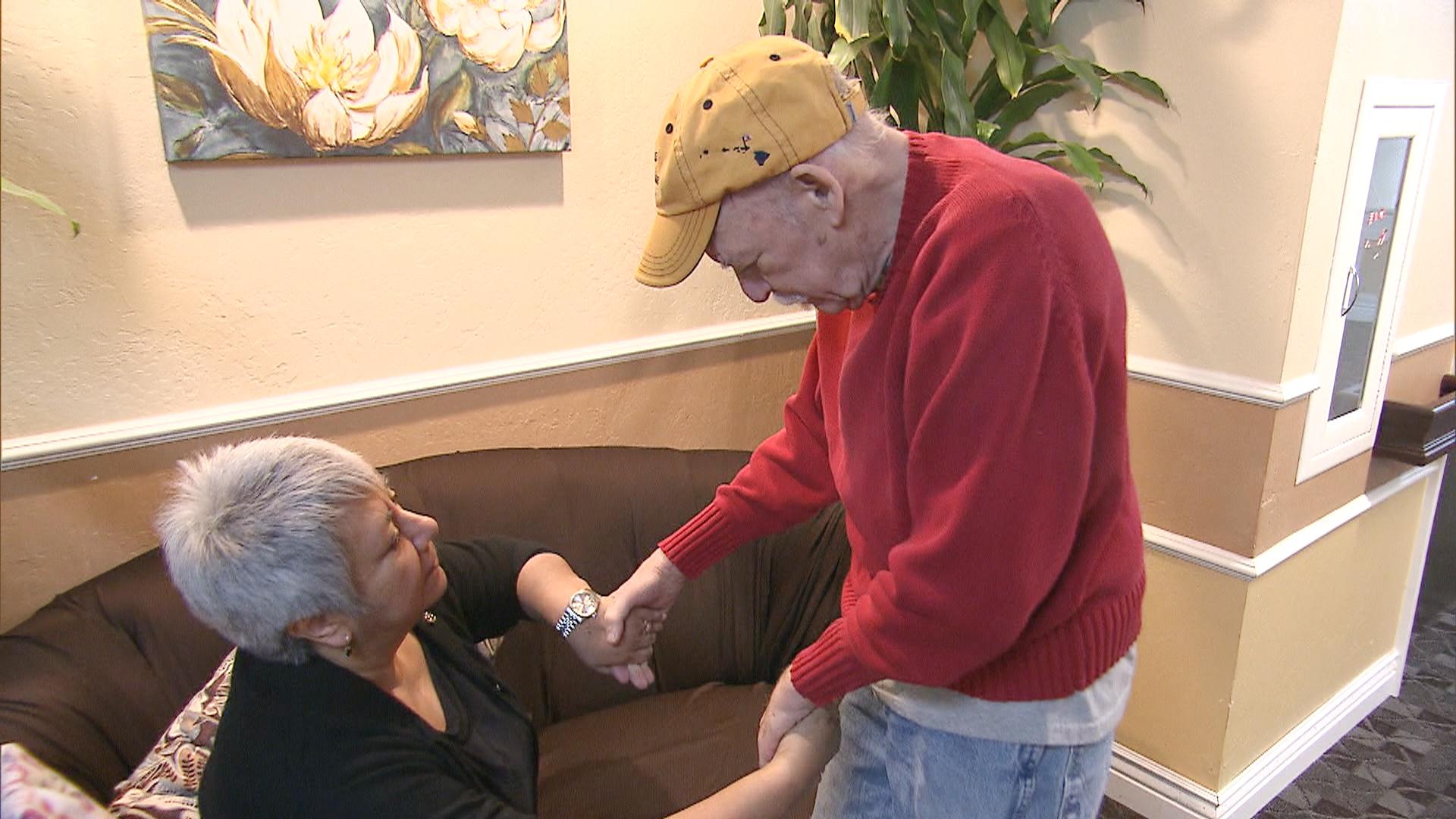
She said physical activity, regular check-ups and abstaining from smoking and drinking were very important for every human to avoid Alzheimer’s. “It is important, as in the initial stages, the patients themselves do not realise that they are suffering from memory loss and very often consider it a minor problem,” she said.

According to scientific estimates published worldwide, a new case of Alzheimer’s arises every four seconds in the world, with the number of people with it set to double every 20 years. The studies also say that by 2040, over 82 million elderly people are expected to have Alzheimer’s if the current numbers hold and no preventive treatment become available.

Renu Vohra, member secretary of ARDSI’s Delhi chapter, said: “A brain-healthy diet is one that reduces the risk of heart disorders and keeps a check on diabetes. The intake of fish oils and folate reduces the dementia risk.” “Miling with friends and family members and having a large social network boost the emotional quotient. Stress clogs the mind and makes it virtually inactive, while the support system provides the healing and keeps one upbeat, at individual one should train the mind to stay calm and positive,” Vohra said.

Source: business standard


Ginger – The Amazing Healing Herb

Ginger is a very good antioxidant and has anti-bacterial, antiseptic, sedative and antipyretic effect. Fresh ginger submerged in warm water, deeply penetrates the tissue, brakes down and melts accumulated mucus, initiate circulation, relieves pain, inflammation and swellings, accelerates detoxification it is very useful in the case of muscle inflammation and chronic back pain.

Ginger – The Amazing Healing Herb

Melts fatty deposits

This healing root speeds up the muscle metabolism, lowers cholesterol, helps in secretion of saliva and ingested food starch, reduces bloating and constipation, all of which contributes to weight loss.
If you want to loose weight fast, add to yo meals fresh or dried ginger or drink tea made from this beneficial root.

Improves mental capacities

Ginger relieves headaches and contribute to a better brain function. If it in any way we use it during the day, we will be fresher and will have more strength and energy, and if in the evening we eat hot vegetable soup with an addition of ginger, we will provide our self a good rest and sleep.

Lowers Stress

Ginger is rich in antioxidant, which helps in harmful chemicals removal, that our body produces when we are under worries, and thus affects the reduction of psychological stress. During this depression treatment it also impairs and the digestion process followed by occurrence of nausea, for which ginger is the ideal cure for these problems too.

Fights against colds and flu

If you regularly use ginger, you can easily prevent and fight the flu or colds, as well as the potential complications from these diseases. Ginger will strengthen our immune system, facilitate breathing and relieve headaches. It encourages sweating, so that`s why it`s excellent for lowering high body temperatures.

Cures Arthritis

Since ginger has anti-inflammatory properties, it is highly effective in the treatment of rheumatoid arthiritis, osteoarthritis and other diseases of the musculoskeletal and connective tissue. Some studies show that in the case of arthritis, ginger is even more effective than other conventional drugs and medications.

Cure for cold hands and feet

Herb ginger is a powerful tool for engaging of the circulatory system.
If you suffer from cold hands and feet you should drink tea from this medicinal herb.

Source: only pure nature


Spatial attention skills don’t seem to decline over time

At least one part of an older person’s brain can still process information as well as younger people, according to new research.

Researchers compared the spatial attention skills of 60 older adults and younger people. Spatial attention is important for many areas of life, from walking and driving to picking up and using items.

Spatial attention skills don't seem to decline over time

“Our studies have found that older and younger adults perform in a similar way on a range of visual and non-visual tasks that measure spatial attention,” Dr. Joanna Brooks, who conducted the experiments as a visiting research fellow at the University of Adelaide in Australia, said in a university news release.

“Both younger (aged 18 to 38 years) and older (55 to 95 years) adults had the same responses for spatial attention tasks involving touch, sight or sound,” noted Brooks, who is now a research fellow in healthy aging at the Australian National University.

The findings were presented at a recent conference in Australia organized by the Australasian Cognitive Neuroscience Society.

“When we think of aging, we think not just of the physical aspects but also the cognitive [mental] side of it, especially when it comes to issues such as reaction time, which is typically slower among older adults. However, our research suggests that certain types of cognitive systems in the right cerebral hemisphere — like spatial attention — are ‘encapsulated’ and may be protected from aging,” Brooks said.

The results challenge current thinking, she said. “We now need to better understand how and why some areas of the brain seem to be more affected by aging than others,” she added.

This type of research could also improve understanding of how diseases such as Alzheimer’s affect the brain, the researchers said.

Data and conclusions presented at meetings are typically considered preliminary until published in a peer-reviewed medical journal.

Source: web md


Heart exercises protect your brain too

Daily exercises to maintain good cardiovascular health also benefits brain, protecting us from cognitive impairment as we age, according to a promising study.

Researchers found that healthy lifestyle helps maintain the elasticity of arteries, thereby preventing downstream cerebrovascular damage and resulting in preserved cognitive abilities in later life.

Our body’s arteries stiffen with age and the vessel hardening is believed to begin in the aorta – the main vessel coming out of the heart before reaching the brain.

Heart exercises protect your brain too

“Indeed, the hardening may contribute to cognitive changes that occur during a similar time frame,” said lead researcher Claudine Gauthier from University of Montreal, Canada.

They found that older adults whose aortas were in a better condition and who had greater aerobic fitness performed better on a cognitive test.

“We think that the preservation of vessel elasticity may be one of the mechanisms that enables exercise to slow cognitive aging,” Gauthier added.

For the study, they worked with 31 young people between ages 18 and 30 and 54 older participants aged between 55 and 75.

This enabled the team to compare the older participants within their peer group and against the younger group who obviously have not begun the ageing processes in question.

The results demonstrated age-related declines in executive function, aortic elasticity and cardiorespiratory fitness, a link between vascular health and brain function, and a positive association between aerobic fitness and brain function.

Source: Times of India


Parkinson’s boosts creativity: Study

Parkinson's boosts creativity


If you are in a creative profession, Parkinson’s may be a blessing in disguise as researchers have found that patients of the nerve cells disease in the area of brain are more creative than their healthy peers.

Those Parkinson’s patients taking higher doses of medication are more artistic than their less-medicated counterparts, the study added.

“It began with my observation that Parkinson’s patients have a special interest in art and have creative hobbies incompatible with their physical limitations,” said Rivka Inzelberg, professor at Tel Aviv University in Israel.

For the study, the researchers conducted tests on 27 Parkinson’s patients treated with anti-Parkinson’s drugs and 27 age and education-matched healthy controls.

The tests included the Verbal Fluency exam, in which a person is asked to mention as many different words beginning with a certain letter and in a certain category (fruit, for example) as possible.

The participants were then asked to undergo a more challenging Remote Association Test, in which they had to name a fourth word (following three given words) within a fixed context.

The groups also took the Tel Aviv University Creativity Test, which tested their interpretation of abstract images and assessed the imagination inherent in answers to questions like “What can you do with sandals?”

The final exam was a version of the Test for a Novel Metaphor, adapted specifically for the study.

Throughout the testing, Parkinson’s patients offered more original answers and more thoughtful interpretations than their healthier counterparts.

In order to rule out the possibility that the creative process evident in the hobbies of patients was linked to obsessive compulsions like gambling and hoarding, to which many Parkinson’s patients fall prey, participants were also asked to fill out an extensive questionnaire.

An analysis indicated no correlation between compulsive behaviour and elevated creativity.

The study appeared in the journal Annals of Neurology.

Source: Business Standard


Now check your eyes, sense of smell to spot Alzheimer’s early

Now check your eyes, sense of smell to spot Alzheimer's early

New research has revealed that Alzheimer’s key biomarker could be identified in the retina and lens of the eye, moreover, the ability to identify odors might indicate the development of cognitive impairment to Alzheimer’s disease in its early ages.

The study found that the decreased ability to identify odors was significantly associated with loss of brain cell function and progression to Alzheimer’s disease. In two other studies, the level of beta-amyloid detected in the eye was significantly correlated with the burden of beta-amyloid in the brain and allowed researchers to accurately identify the people with Alzheimer’s in the studies.

Beta-amyloid protein is the primary material found in the sticky brain “plaques” characteristic of Alzheimer’s disease. It was known to build up in the brain many years before typical Alzheimer’s symptoms of memory loss and other cognitive problems.

Heather Snyder, Ph.D., said that advanced researches were needed in the very promising area of Alzheimer’s biomarkers because early detection was essential for early intervention and prevention, when new treatments become available.

Clinically, at this time it was only possible to detect Alzheimer’s late in its development, when significant brain damage has already occurred, however, biological markers of Alzheimer’s disease might be able to detect it at an earlier stage.

Source: Business standard


Brain signal that may help in drug de-addiction

braintorm

Researchers have discovered a new form of neurotransmission that influences long-lasting memory created by addictive drugs like cocaine and opioids and the craving for them.

Loss of this type of neurotransmission creates changes in brain cells that resemble the changes caused by drug addiction.

The findings suggest that targeting this type of neurotransmission might lead to new therapies for treating drug addiction.

“Molecular therapies for drug addiction are pretty much non-existent,” said Collin Kreple from the University of Iowa in the US.

“I think this finding at least provides the possibility of a new molecular target,” Kreple added.

This neurotransmission involves proteins called acid-sensing ion channels (ASICs), which have previously been shown to promote learning and memory, and which are abundant in a part of the brain involved in drug addiction.
The experiments showed that loss of ASIC signalling increases learned drug-seeking in mice.

When mice learned to associate one side of a chamber with receiving cocaine, animals that lacked the ASIC protein developed an even stronger preference for the “cocaine side” than control mice, suggesting that loss of ASIC had increased addiction behaviour.

The same result was seen for morphine, which has a different mechanism of action than cocaine.

In a second experiment, rats learned to press a lever to self-administer cocaine. Blocking or removing ASIC in the rat brains caused the animals to self-administer more cocaine than control animals.

Conversely, increasing the amount of ASIC by over-expressing the protein seemed to decrease the animals’ craving for cocaine.

The study was published in the journal Nature Neuroscience.

Source: yahoo news


3D brain view may help treat Alzheimer’s, Parkinson’s

3D brain view may help treat Alzheimer's Parkinson's

In a breakthrough that may help in developing drugs for Alzheimer’s and other neurological disorders, researchers have developed a 3D view of an important receptor in the brain.

This receptor allows us to learn and remember, and its dysfunction can result in a wide range of neurological diseases including Alzheimer’s, Parkinson’s, schizophrenia and depression.

The unprecedented view gives scientists new insight into how the receptor – called the NMDA receptor – is structured.

And importantly, the new detailed view gives vital clues for developing drugs to combat neurological diseases and conditions.

“This is the most exciting moment of my career,” said Eric Gouaux, a senior scientist with Oregon Health and Science University in the US.

“The NMDA receptor is one of the most essential, and still sometimes mysterious, receptors in our brain. Now, with this work, we can see it in fascinating detail,” he said.

Receptors facilitate chemical and electrical signals between neurons in the brain allowing them to communicate with each other.

The NMDA (N-methyl-D-aspartate) receptor facilitates neuron communication that is the foundation of memory, learning and thought.

Malfunction of the NMDA receptor occurs when it is increasingly or decreasingly active.

The NMDA receptor makeup includes receptor “subunits” – all of which have distinct properties and act in distinct ways in the brain, sometimes causing neurological problems.

Prior to Gouaux’s study, scientists had only a limited view of how those subtypes were arranged in the NMDA receptor complex and how they interacted to carry out specific functions within the brain and the central nervous system.

Gouaux’s team of scientists created a 3D model of the NMDA receptor through a process called X-ray crystallography.

“This new detailed view will be invaluable as we try to develop drugs that might work on specific subunits and therefore help fight or cure some of these neurological diseases and conditions,” Gouaux said.

“Seeing the structure in more detail can unlock some of its secrets and may help a lot of people,” he added.

The findings were published online in the journal Nature.

Source: Hindustan Times


Now, a ‘smart cap’ to map brain functions

640_electroencephalograph

The functional magnetic resonance imaging (MRI), most commonly used in brain mapping method, may not be ideal for children and patients with implanted electrical devices such as pacemakers, cochlear implants and deep brain stimulators. The magnetic fields used in the method may disrupt either the function or safety of these devices.

Researchers have now developed a “smart cap”, a new brain scanning system that shines tiny lights onto the head and works just as well as magnetic brain scanner.

The instrument uses a technology called diffuse optical tomography (DOT) and works by detecting light transmitted through the head and capturing the dynamic changes in the colours of the brain tissue.

The new DOT instrument covers two-thirds of the head and for the first time can image brain processes taking place in multiple regions and brain networks.

“When the neuronal activity of a region in the brain increases, highly oxygenated blood flows to the parts of the brain doing more work and we can detect that,” Joseph Culver from Washington University was quoted as saying.

The researchers validated the performance of DOT by comparing its results to fMRI scans, Daily Mail reported.

The study appeared in the journal Nature Photonics.

Source: chennai online


Fully-formed teeth growing inside baby’s BRAIN: Medical Miracle

A four-month-old baby boy was found to have teeth growing in his brain. The unnamed infant in Maryland, U.S., had a rare type of brain tumour which contained multiple fully formed teeth.

He is thought to be the first person in the world to be found to have teeth within this type of brain tumour. The baby required surgery to remove the tumour, but, a year on, he is now making a good recovery.

The child first came to the attention of doctors after a routine health check revealed his head was growing faster than expected.  According to a report in the New England Journal of Medicine, he had an MRI scan which revealed a tumour near his pituitary gland which measured 4.1cm by 4cm by 3.5cm.

The scan revealed that the tumour also contained teeth very similar to those found in the lower jaw. The baby underwent surgery to remove the tumour and further tests revealed it was a slow-growing mass called adamantinomatous craniopharyngioma.

The tumour is thought to have arisen from Rathke’s pouch, an embryonic precursor to part of the pituitary gland. These tumours are sometimes filled with viscous, yellow fluid containing cholesterol crystals.

They usually start in the area around the pituitary gland but spread into surrounding areas and they often recur after they have, seemingly, been completely removed. It is very rare for them to spread outside the brain.

It is now a year since the body underwent surgery and he is making good progress. However, he has had to have a shunt fitted to drain brain and spinal fluid from his head. He also has to take thyroid and adrenal hormone-replacement drugs.

He still undergoes regular MRI scans to ensure the tumour has not returned. The doctors who treat the boy say it has long been suspected that the type of tumour he suffered from form from the same cells as those involved in the creation of teeth.

However, until now, surgeons had never actually seen teeth within one of these tumours. Dr Narlin Beaty, a neurosurgeon at the University of Maryland Medical Center, who performed the boy’s surgery, told Live Science: ‘It’s not every day you see teeth in any type of tumour in the brain. In a craniopharyngioma, it’s unheard of.’

He added that these tumours often contain some calcium deposits ‘but when we pulled out a full tooth…I think that’s something slightly different’.

Source: Daily mail