Stem cells may help cure bladder issues

Scientists have now managed to produce tissue from human stem cells that could be transplanted into patients with defective or diseased bladder, says a study.

For the first time, scientists have succeeded in coaxing laboratory cultures of human stem cells to develop into the specialized, unique cells needed to repair a patient’s defective or diseased bladder.

The breakthrough was developed at the University of California’s (UC) Davis Institute for Regenerative Cures and published in the scientific journal Stem Cells Translational Medicine.

It is significant because it provides a pathway to regenerate replacement bladder tissue for patients whose bladders are too small or do not function properly, such as children with spina bifida and adults with spinal cord injuries or bladder cancer, reported Science Daily.

“Our goal is to use human stem cells to regenerate tissue in the lab that can be transplanted into patients to augment or replace their malfunctioning bladders,” said Eric Kurzrock, professor and head of the division of paediatric urologic surgery at UC Davis Children’s Hospital and lead scientist of the study.

Another benefit of the UC Davis study is the insight it may provide about the pathways of bladder cancer, which is diagnosed in more than 70,000 Americans each year, according to the National Cancer Institute.

Source: business standard


New lens design improves kidney stone treatment

Duke engineers have devised a way to improve the efficiency of lithotripsy — the demolition of kidney stones using focused shock waves. After decades of research, all it took was cutting a groove near the perimeter of the shock wave-focusing lens and changing its curvature.I’ve spent more than 20 years investigating the physics and engineering aspects of shock wave lithotripsy,” said Pei Zhong, the Anderson-Rupp Professor of Mechanical Engineering and Materials Science at Duke University. “And now, thanks to the willingness of Siemens (a leading lithotripter manufacturer) to collaborate, we’ve developed a solution that is simple, cost-effective and reliable that can be quickly implemented on their machines.”

The study appears in the Proceedings of the National Academy of Sciences.

The incidence of kidney stones in the United States has more than doubled during the past two decades, due at least in part to the expanding waistlines of its citizens. The condition has also been linked to hot, humid climates and high levels of stress—a combination of living environments that seems to have led to a rise in kidney stone rates of veterans returning home from Iraq and Afghanistan.

During the past two decades, lithotripter manufacturers introduced multiple changes to their machines. Rather than having patients submerged in a bath of lukewarm water, newer machines feature a water-filled pouch that transfers the shock wave into the flesh. An electrohydraulic shock wave generator used in the past was replaced by an electromagnetic model that is more powerful, more reliable and more consistent.

The new designs made the devices more convenient and comfortable to use, but reduced the effectiveness of the treatment. After years of research, Zhong and his colleagues have determined why.

The increased power in some third-generation shock wave lithotripters narrowed the wave’s focal width to reduce damage to surrounding tissues. But this power jump also shifted the shock wave’s focal point as much as 20 millimetres toward the device, ironically contributing to efficiency loss and raising the potential for tissue damage. The new electromagnetic shock wave generators also produced a secondary compressive wave that disrupted one of the primary stone-smashing mechanisms, cavitation bubbles.

“We were presented with the challenge of engineering a design solution that mitigated these drawbacks without being too expensive,” said Zhong. “It had to be something that was effective and reliable, but also something that the manufacturer was willing to adopt. So we decided to focus on a new lens design while keeping everything else in their system intact.”

The solution was to cut a groove near the perimeter of the backside of the lens and change its geometry. This realigned the device’s focal point and optimized the pressure distribution with a broad focal width and lower peak pressure. It also allowed more cavitation bubbles to form around the targeted stone instead of in the surrounding tissue.

In laboratory tests, the researchers sent shock waves through a tank of water and used a fibre optic pressure sensor to ensure the shock wave was focusing on target. They broke apart synthetic stones in a model human kidney and in anaesthesized pigs and used a high-speed camera to watch the distribution of cavitation bubbles forming and collapsing—a process that happens too fast for the human eye to see.

The results showed that while the current commercial version reduced 54 per cent of the stones into fragments less than two millimetres in diameter, the new version pulverized 89 per cent of the stones while also reducing the amount of damage to surrounding tissue. Smaller fragments are more easily passed out of the body and less likely to recur.

“We feel we have exceeded expectations in our evaluation of this new lens design, which is based on solid physics and engineering principles,” said Zhong, who expects the new lens to enter clinical trials in Germany this summer.

“My hope is that this will be a breaking point demonstrating that effective, interactive collaboration between academia and industry can really improve the design of lithotripters that will benefit millions of stone patients worldwide who suffer from this painful disease,” Zhong said.

“Our design, in principle, can be adapted by other manufacturers to improve their machines as well. I would like to see all lithotripsy machines improved so that urologists can treat stones more effectively and patients can receive better treatment and feel more comfortable with the procedure,” he added.

Source: India Medical Times


Cerebrospinal fluid test may detect Alzheimer’s

Researchers at the University of Texas, in Houston, analysed CSF samples from 50 people with Alzheimer’s disease, 37 people with other neurodegenerative diseases such as Parkinson’s disease, and 39 people with other brain diseases who had normal memory and thinking abilities. They set out to discover whether small fragments of the protein amyloid, which accumulates into clumps in the brain during Alzheimer’s, were present in the CSF of people with the disease.

Their results showed more of these fragments of protein in the CSF of people with Alzheimer’s compared to those with other diseases. The researchers suggest that more research is needed to explore whether the method could detect the disease at its earliest stages.

Dr Simon Ridley, Head of Research at Alzheimer’s Research UK, the UK’s leading dementia research charity, said:
“This very small study suggests a potential way to identify people with Alzheimer’s disease, but it’s too early to tell how accurate this method might be or whether it can distinguish people with the disease from healthy people. We need to see further research in much larger groups of people before we can know whether this approach will be successful.

“Diagnosing Alzheimer’s can be difficult, and the ability to give an accurate diagnosis would be a real boost, allowing people access to the right care and existing treatments. The ability to detect Alzheimer’s accurately is also a key goal for research, as it’s important to be able to test potential new treatments in the right people. Investment in research is vital to develop better diagnosis tools and, crucially, for better treatments to be made available to those who desperately need them.”

Source: alzheimers Research


Shift workers beware: Sleep loss may cause brain damage, new research says

Are you a truck driver or shift worker planning to catch up on some sleep this weekend? Cramming in extra hours of shut-eye may not make up for those lost pulling all-nighters, new research indicates.

The damage may already be done — brain damage, that is, said neuroscientist Sigrid Veasey from the University of Pennsylvania.

Alzheimer’s & Sleep

The widely held idea that you can pay back a sizeable “sleep debt” with long naps later on seems to be a myth, she said in a study published this week in the Journal of Neuroscience.

Long-term sleep deprivation saps the brain of power even after days of recovery sleep, Veasey said. And that could be a sign of lasting brain injury. Veasey and her colleagues at the University of Pennsylvania medical school wanted to find out, so, they put laboratory mice on a wonky sleep schedule that mirrors that of shift workers.

They let them snooze, then woke them up for short periods and for long ones.
Then the scientists looked at their brains — more specifically, at a bundle of nerve cells they say is associated with alertness and cognitive function, the locus coeruleus.

They found damage and lots of it.
“The mice lose 25% of these neurons,” Veasey said.
This is how the scientists think it happened.
When the mice lost a little sleep, nerve cells reacted by making more of a protein, called sirtuin type 3, to energize and protect them.

But when losing sleep became a habit, that reaction shut down. After just a few days of “shift work” sleep, the cells start dying off at an accelerated pace.
The discovery that long-term sleep loss can result in a loss of brain cells is a first, Veasey said.

“No one really thought that the brain could be irreversibly injured from sleep loss,” she said. That has now changed.

More work needs to be done on humans, she said. And her group is planning to study deceased shift workers to see if they have the same kind of nerve damage. They hope their research will result in medicines that will help people working odd hours cope with the consequences of irregular sleep.

Source: CNN


ADHD treatment may increase obesity risk

Previous studies have linked ADHD with an increased risk of obesity. Now researchers at the Johns Hopkins Bloomberg School of Public Health say the reason may be because of ADHD stimulant medications, rather than the condition itself.

Using the electronic health record data from the Gesinger Health System, the researchers analyzed 163,820 children between the ages of 3 to 18 years. They found that children who had ADHD and were treated with stimulants experienced a fast adolescent BMI growth compared to children with no ADHD history or stimulant use. Also, the earlier children began using stimulants, the stronger these weight effects were.

This research, published in the journal Pediatrics, backs current research indicating a relationship between ADHD and obesity. However, more research is needed to determine the exact reason behind this connection.

Source: health central


3,000-year-old skeleton with cancer helps scientists understand disease evolution

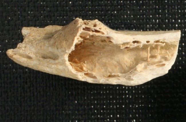
A research team from Europe recently discovered a 3000 year old skeleton, which was believed to be a young adult male, inside in a tomb in Sudan, and it showed evidence that the person had metastatic carcinoma. The cancer had spread to various parts of the body, so this made it the oldest cancer sample that had ever been found, stated a Durham University press release.

Cancer had always been thought of as a present day condition and it seemed to have been basically non-existent during most of human history, despite being one of the top causes of death in modern times. The skeleton underwent investigation via radiography and also electron microscope scanning. By using these cutting-edge methods, the scientists were able to find that the skeleton was riddled with lesions and tumors that covered the collar bones, upper and lower arms, shoulder blades, ribs, vertebrae, thigh bones and the pelvis.

The findings might be able to aid researchers receive awareness into what was the cause of cancer in the past and how the disease has evolved. Scientists plan on using DNA studies of ancient remains to try and find cancer mutations. Records of cancer from the past have been extremely uncommon and medical researchers think that the modern way of life is behind why the number of cancer cases has risen so radically.

The cancer might have been caused from environmental pollutants such as parasites and wood fires and parasites, or it may have been inherited. It is also possible that some sort of schistosomiasis infection could have been a trigger, as this can cause bladder cancer and is known to be widespread in that area from about 15,000 B.C.

There is very little known about the evolution of cancer in ancient human populations apart from a few written references and a tiny number of skeletons that have cancer signs. Any understandings that scientists can gather from ancient human remains such as the ones found aid in the understanding of the history and evolution of modern diseases. The researches stated that the smaller lesions located on the bones could only have been caused by some sort of cancer from soft tissues in the body even though the precise origin was impossible to figure out from only the bones.

The male skeleton was thought to have been between the ages of 24 and 35 when he died. After this he was buried inside a painted sarcophagus. Before this discovery, there were only two other examples of possible metastatic cancer and they each dated much younger than this skeleton. Scientists believe that by taking an evolutionary line of attack to cancer, information that is discovered from ancient human remains might provide vital elements in finding different ways to look at one of the world’s biggest health problems.

The World Health Organization states that cancer takes the lives of nearly 14 million individuals each year. It is the hope of many researchers that these brand new findings are able to help scientists disentangle the primary causes of cancer so that the disease can be better treated and also stopped. Insights that are found from such remains really help researchers understand the evolution and history of modern diseases.

The research team found the skeleton, which was believed to be a young adult male, inside in a tomb in Sudan, and it showed evidence that the person had metastatic carcinoma. The cancer had spread to various parts of the body, so this made it the oldest cancer sample that had ever been found, stated a Durham University press release.

Source: Liberty voice


Colon cancer rates declining in older Americans

Colon cancer incidence rates have dropped 30 percent in the U.S. in the last 10 years among adults 50 and older due to the widespread uptake of colonoscopy, with the largest decrease in people over age 65. Colonoscopy use has almost tripled among adults ages 50 to 75, from 19 percent in 2000 to 55 percent in 2010.

The findings come from Colorectal Cancer Statistics, 2014, published in the March/April issue of CA: A Cancer Journal for Clinicians. The article and its companion report, Colorectal Cancer Facts & Figures, were released today by American Cancer Society researchers as part of a new initiative by the National Colorectal Cancer Roundtable to increase screening rates to 80 percent by 2018.

Colorectal cancer, commonly called colon cancer, is the third most common cancer and the third leading cause of cancer death in men and women in the United States. Its slow growth from precancerous polyp to invasive cancer provides a rare opportunity to prevent cancer through the detection and removal of precancerous growths. Screening also allows early detection of cancer, when treatment is more successful. As a result, screening reduces colorectal cancer mortality both by decreasing the incidence of disease and by increasing the likelihood of survival.

Using incidence data from the National Cancer Institute’s Surveillance, Epidemiology, and End Results (SEER) program and the Centers for Disease Control and Prevention’s National Program of Cancer Registries, as provided by the North American Association of Central Cancer Registries (NAACCR), researchers led by Rebecca Siegel, MPH found that during the most recent decade of data (2001 to 2010), overall incidence rates decreased by an average of 3.4 percent per year. However, trends vary substantially by age. Rates declined by 3.9 percent per year among adults aged 50 years and older, but increased by 1.1 percent per year among men and women younger than 50. That increase was confined to tumors in the distal colon and rectum, patterns for which a rise in obesity and emergence of unfavorable dietary patterns has been implicated.

Most strikingly, the rate of decline has surged among those 65 and older, with the decline accelerating from 3.6 percent per year during 2001-2008 to 7.2 percent per year during 2008-2010. The “larger declines among Medicare-eligible seniors likely reflect higher rates of screening because of universal insurance coverage,” the authors write. “In 2010, 55 percent of adults aged 50 to 64 years reported having undergone a recent colorectal cancer screening test, compared with 64 percent of those aged 65 years and older.”

Like incidence, mortality rates have also declined most rapidly within the past decade. From 2001 to 2010, rates decreased by approximately 3 percent per year in both men and women, compared with declines of approximately 2 percent per year during the 1990s.

“These continuing drops in incidence and mortality show the lifesaving potential of colon cancer screening; a potential that an estimated 23 million Americans between ages 50 and 75 are not benefiting from because they are not up to date on screening,” said Richard C. Wender, M.D., American Cancer Society chief cancer control officer. “Sustaining this hopeful trend will require concrete efforts to make sure all patients, particularly those who are economically disenfranchised, have access to screening and to the best care available.”

The data is being released at the launch of a nationwide effort to increase colorectal cancer screening rates to 80% by 2018. Public health leaders, including Assistant Secretary for Health Howard Koh, MD, MPH and American Cancer Society CEO, John R. Seffrin, PhD will join dozens of members of the National Colorectal Cancer Roundtable (NCCRT) at the National Press Club in Washington, D.C. on March 17th at 1:00 PM EDT. The NCCRT, an organization co-founded by the American Cancer Society and the U. S. Centers for Disease Control and Prevention, will focus on dramatically increasing colorectal cancer screening rates in the U.S. over the next four years, and increasing awareness of the potential for early detection and prevention of this cancer.

Source: Science daily


New vaginal gel could help protect women against HIV

Researchers have revealed that a new after-sex vagina gel can be used by women to protect themselves against HIV.

According to scientists at Centers for Disease Control and Prevention in Atlanta, Georgia, drugs applied three hours after exposure to the virus could protect female monkeys from a type of HIV, the BBC reported.

that this study would require large clinical trials to test any new treatment and that condoms still remain the best defense against HIV.

Scientists found that the gel protected five out of six monkeys from an animal-human laboratory strain of HIV, when it was applied before or three hours after infection.

Dr Charles Dobard, of the division of HIV/Aids prevention, said that the gel used is a promising after-sex vaginal gel to prevent HIV infection and studies still need to look for the window of opportunity.

Dr Andrew Freedman, reader and consultant in infectious diseases at Cardiff University School of Medicine, said that the gel contained a different class of anti- HIV drug, which attacks the virus at a later stage in infection.

The study is published in the journal Science Translational Medicine.

Source: Zee news


Japanese scientist withdraws ‘groundbreaking’ stem cell research

A Japanese scientist has asked for the findings of his groundbreaking study in stem cells to be withdrawn amid doubts over its quality.

According to Reuters news agency, Prof Teruhiko Wakayama of the University of Yamanashi told Japanese TV that when he was conducting the experiment, he believed that it was absolutely right, but many mistakes have emerged which has led him to withdraw the research paper, the BBC reported.

Several questions have been raised about the images used in the scientific report which claimed that dipping skin cells in acid could cheaply and quickly convert them into stem cells.

The original study, which was published in the journal Nature, had found that stem cells no longer needed to be taken from embryos or made by complicated and costly genetic tampering, and had been hailed as “remarkable” and as a “major scientific discovery”.

Source: Business Standard


Doctors grow ears, noses using body fat stem cells

British scientists are aiming to grow ears and noses in a laboratory to transplant then into humans.

Scientists from Great Ormond Street Hospital and University College London have managed to use abdominal body fat and turn it into cartilage. It is now hoped that the technique could help patients who have been born with microtia, which means the ear fails to develop properly, or who have been in an accident.

At the moment, surgeons take cartilage from other parts of the body to treat children with facial defects. The painful procedure sees them shape the nose or ear, and implant it into the child

The new technique would mean that doctors ‘grow’ the organ separately using a tiny sample of fat from the child. Stem cells would be extracted and grown from it. Scientists would place an ear-shaped ‘nano-scaffold’ into the stem cell broth so that they take on the correct shape and structure.

This would then be placed beneath the skin. Although it would not help with hearing it would be biologically the same as the real thing, the Telegraph reports.  The breakthrough has been published in the journal Nanomedicine: Nanotechnology, Biology and Medicine.  Neil Bulstrode, consultant plastic surgeon at Great Ormond Street Hospital, co-authored the research.

He said: ‘It is such an exciting prospect. If we could produce a block of cartilage using stem cells and tissue engineering, this would be the holy grail for our field.’ Each year thousands of children are born with a congenital deformity called microtia, when the external ear is not fully developed. Many have an intact inner ear, but experience hearing loss due to the missing external structure.

But the research could also have implications for the future of other transplants, and could be used to create bone and other tissue. The report said the technique procedure could ‘help to improve stability, integration and functionality of engineered transplants while avoiding tissue rejection’.

Dr Patrizia Ferretti, the head of developmental biology at UCL, said it would be useful for children because it means there is no need for immune suppression.

She said: ‘At the moment we take cartilage out of the ribs which means a major additional surgical procedure that creates a permanent defect, as the rib cartilage does not regrow.
‘But with this technique you could seed the stem cells on to a mould of a healthy ear, or use 3D printing to make the ear shaped scaffold-containing cells that can then be turned into cartilage.’

Source: Daily mail