Finland researchers develop handheld camera for early detection of skin cancer

VTT Technical Research Centre of Finland has developed a lightweight, handheld, ultra-precision hyperspectral camera for the detection of skin cancers and their precursors.

From the surface of the skin, the camera recognises early stages of cancer that are invisible to the naked eye.

Collaborators in the pilot study are the University of Jyvaskyla, the Paijat-Hame Central Hospital and the Skin and Allergy Hospital of Helsinki University Central Hospital. The preliminary results are promising, say the researchers.

The hand-held, mobile hyperspectral camera images the skin region in two seconds. The large field of view (12 cm2) enables the detection of large skin areas at once.

In the pilot study, the camera has been used to detect the skin areas with field cancerization i.e. areas of multiple skin cancer precursors, actinic keratoses, for early treatment of the affected areas.

The hyperspectral camera has also been used to detect the borders of poorly delineated skin tumours, such as lentigo malignas, which are difficult to detect by the naked eye, in order to avoid the need for re-excisions.

Developed by VTT on the basis of the Fabry-Perot interferometer, the hyperspectral camera captures images in up to 70 narrow wavelengths, whereas a regular camera uses only three.

The spectral image generated is a three-dimensional cube built of numerous layers of greyscale images, each of which has been taken within a limited wavelength range.

A spectrum for each pixel of the spectral image is formed by the images within the cube. Different biological tissues can be identified by their reflected spectra in hyperspectral images.

Computational methods are used to interpret these images, in order to determine the position and size of the tumour to be treated. In the ongoing pilot study, all results are being verified by histopathological sampling.

Patents have been granted for the hyperspectral camera in the US and in Finland. Heikki Saari, principal scientist at VTT, is the inventor of this patented device.

Skin cancer rates have been growing exponentially, due to population ageing and UV damage caused by excessive exposure to sunlight.

The camera is owned by the University of Jyvaskyla. It can also be used for various applications of a more general nature, according to a statement by VTT.

Source: India Medical Times


When Breast Cancer Spreads

If your cancer spreads beyond your breast and the nearby lymph nodes, it’s called advanced cancer, or metastatic cancer. The most common places it spreads to are the liver, lungs, bones, and brain.

News that your cancer has spread is scary, but there are many treatments that work for metastatic breast cancer.

“The majority of women with metastatic breast cancer can move forward with their therapies while continuing their regular lifestyle — working, taking care of their families, exercising, and traveling,” says Erica L. Mayer, MD, MPH, of the Dana-Farber Cancer Institute in Boston.

“We often think of metastatic breast cancer as a chronic disease, like diabetes,” says Mayer. A Different Treatment Schedule

Treatments for advanced breast cancer may go on without an end date, to keep the cancer under control. You’ll visit the clinic on a regular basis and you’ll get to know the health care team.

“If the treatment works, you’ll stay on it as long as it’s working well without side effects,” says Rita Nanda, MD, of the University of Chicago’s breast cancer program. If not, your doctor will try different treatments.

Your doctor is likely to suggest chemotherapy because it travels through your entire body. “Metastatic breast cancer is a whole-body disease,” Mayer says.

You may also need hormone therapy. Targeted drugs are another option. They work directly on the changes within cancer cells. These combinations can make chemotherapy work better.

Sometimes surgery or radiation can help ease symptoms. Regular Tests Keep Tabs on Your Cancer

Occasionally, you’ll have imaging tests to see how treatments are working and whether the cancer has spread. Common imaging tests include:

CT scans, where an X-ray machine circles around as you lie on a table Bone scans with an injection that helps show areas with cancer (scintigraphy) PET scans with a special camera and a tracer chemical that goes in your arm by IV
“CT scans examine the chest and abdomen,” says Richard J. Bleicher, MD, of the Fox Chase Cancer Center in Philadelphia. “You can see something on organs like the liver or sometimes the bones.” Sometimes results are combined for a PET CT scan. A computer merges the images to find hot spots that may be cancer.

Your doctor will tell you how often you need these tests, based on the stage of your cancer.

Source: Web md


MIT engineers develop paper diagnostic for cancer

Cancer rates in developing nations have climbed sharply in recent years, and now account for 70 per cent of cancer mortality worldwide. Early detection has been proven to improve outcomes, but screening approaches such as mammograms and colonoscopy, used in the developed world, are too costly to be implemented in settings with little medical infrastructure.

To address this gap, Massachusetts Institute of Technology (MIT) engineers have developed a simple, cheap, paper test that could improve diagnosis rates and help people get treated earlier. The diagnostic, which works much like a pregnancy test, could reveal within minutes, based on a urine sample, whether a person has cancer. This approach has helped detect infectious diseases, and the new technology allows noncommunicable diseases to be detected using the same strategy.

The technology, developed by MIT professor and Howard Hughes Medical Institute investigator Sangeeta Bhatia, relies on nanoparticles that interact with tumour proteins called proteases, each of which can trigger release of hundreds of biomarkers that are then easily detectable in a patient’s urine.

“When we invented this new class of synthetic biomarker, we used a highly specialized instrument to do the analysis,” says Bhatia, the John and Dorothy Wilson Professor of Health Sciences and Technology and Electrical Engineering and Computer Science. “For the developing world, we thought it would be exciting to adapt it instead to a paper test that could be performed on unprocessed samples in a rural setting, without the need for any specialized equipment. The simple readout could even be transmitted to a remote caregiver by a picture on a mobile phone.”

Bhatia, who is also a member of MIT’s Koch Institute for Integrative Cancer Research and Institute for Medical Engineering and Science, is the senior author of a paper describing the particles in the Proceedings of the National Academy of Sciences.

In 2012, Bhatia and colleagues introduced the concept of a synthetic biomarker technology to amplify signals from tumour proteins that would be hard to detect on their own. These proteins, known as matrix metalloproteinases (MMPs), help cancer cells escape their original locations by cutting through proteins of the extracellular matrix, which normally holds cells in place.

The MIT nanoparticles are coated with peptides (short protein fragments) targeted by different MMPs. These particles congregate at tumour sites, where MMPs cleave hundreds of peptides, which accumulate in the kidneys and are excreted in the urine.

In the original version of the technology, these peptides were detected using an instrument called a mass spectrometer, which analyses the molecular makeup of a sample. However, these instruments are not readily available in the developing world, so the researchers adapted the particles so they could be analysed on paper, using an approach known as a lateral flow assay — the same technology used in pregnancy tests.

To create the test strips, the researchers first coated nitrocellulose paper with antibodies that can capture the peptides. Once the peptides are captured, they flow along the strip and are exposed to several invisible test lines made of other antibodies specific to different tags attached to the peptides. If one of these lines becomes visible, it means the target peptide is present in the sample. The technology can also easily be modified to detect multiple types of peptides released by different types or stages of disease.

In tests in mice, the researchers were able to accurately identify colon tumours, as well as blood clots. Bhatia says these tests represent the first step toward a diagnostic device that could someday be useful in human patients.

“This is a new idea — to create an excreted biomarker instead of relying on what the body gives you,” she says. “To prove this approach is really going to be a useful diagnostic, the next step is to test it in patient populations.”

To help make that happen, the research team recently won a grant from MIT’s Deshpande Centre for Technological Innovation to develop a business plan for a startup that could work on commercializing the technology and performing clinical trials.

Bhatia says the technology would likely first be applied to high-risk populations, such as people who have had cancer previously, or had a family member with the disease. Eventually, she would like to see it used for early detection throughout developing nations.

Such technology might also prove useful in the United States, and other countries where more advanced diagnostics are available, as a simple and inexpensive alternative to imaging. “I think it would be great to bring it back to this setting, where point-of-care, image-free cancer detection, whether it’s in your home or in a pharmacy clinic, could really be transformative,” Bhatia says.

With the current version of the technology, patients would first receive an injection of the nanoparticles, then urinate onto the paper test strip. To make the process more convenient, the researchers are now working on a nanoparticle formulation that could be implanted under the skin for longer-term monitoring.

The team is also working to identify signatures of MMPs that could be exploited as biomarkers for other types of cancer, as well as for tumours that have metastasized.

Source: India Medical Times


An adult in the pediatric ward: What the littlest Cancer Avengers taught me

140217161534-little-superheroes-ann-and-andreas-horizontal-gallery

Three years ago, I was diagnosed with a rare pediatric bone cancer called osteosarcoma. In my case, it was super-rare, because I was 43.

I went to see Dr. Paul Meyers at Memorial Sloan Kettering Cancer Center in New York, six hours away from our home in upstate New York. At the hospital, my husband and I followed instructions to the B elevators and got off on the eighth floor.

The doors opened to a brightly colored playroom, funky lounge chairs and really big fish tanks.

Meyers is a pediatric oncologist, and because I had a pediatric cancer (age not withstanding), I would be treated where he worked: in the pediatric cancer center.

Meyers explained, sans sugar-coating, that my cancer was particularly aggressive, and so the treatment would be, too. I explained that I loved my job and my life, and am one tough chick, so I planned on working through it all.

Meyers pressed on. I would endure nine rounds of three types of chemotherapy in a not-so-delicious-chemo cocktail. After three rounds, there would be limb-salvage surgery where they remove my cancerous bones and replace them with titanium. Or amputate.

I should expect to be fully debilitated by this treatment, Meyers said — to be in a wheelchair for more than a year, to stop working at my job that I loved, and to close my company that I had worked hard to build. Long-term damage to my hearing, heart, bladder and extremities because of high doses of chemo were to be expected.

At the time, I was unsure of almost everything, including how I felt about being in the Pediatric Day Hospital as a patient.

We learned the hospital would be my home away from home during my nearly yearlong treatment. I spent at least one week of every month with the sickest people you can imagine. Little people with no hair, missing limbs and treacherous looking scars; it was harrowing at first.

Then I became one of them: No hair. Giant, treacherous scar. Wheelchair. Ever-present IV pole, and dusty-rose colored kidney-shaped bowl to throw up in. These were all outward signs of a fraternity of warriors that no one wants to belong to. They all were enduring the same grueling treatment I was — only they were, on average, 10 years old.

This fraternity of Cancer Avengers was wise in ways beyond their years. When faced with the courage and bravery of these little superheroes, I had to give myself the “Put your big girl pants on” speech more than once.

On my first day of treatment, while I was scrolling through my Facebook feed by the fish tank, two boys next to me started discussing their Make-A-Wish requests. Adam, about 12 years old, had just returned from a rainforest trip and asked what Sam’s wish was going to be. Sam said they couldn’t give him what he wished for. Adam disagreed, enthusiastically conveying that any wish could be granted. Sam stood firm: It was not possible.

Well, what is it that you want anyway? Adam wanted to know. By now, I also wanted to know.

“I want normal,” was Sam’s answer. “I want to go to school and basketball practice, complain about my parents and homework and turn 12.”

Silence from Adam. Silence all around. Even a superhero knows when he is defeated.
I looked down at my phone, trying to distract myself and read through my tears. A Facebook friend was complaining about turning 44. In the moment, it was like complaining about being too rich or having too much food to eat. My friend had been granted 32 more years than this kid dared dream of living. So had I.

Source: BBC


When Men Get Breast Cancer?

That is the message of a provocative new photography series featuring the faces, and scars, of men with breast cancer.

The vast majority of the photos in that project are of young women, shown topless with scars where their breasts used to be. The pictures, which are both shocking and beautiful, are featured in a traveling exhibition that will be on display next month in Toronto.

But most visitors to the Scar Project find the photos on the Internet, where they have been viewed by millions of people. One of those people is Oliver Bogler, a cancer biologist in Houston who found out that he had breast cancer 18 months ago after noticing a lump in his chest.

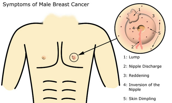
As in a woman’s breast, the duct cells in a man’s breast can undergo cancerous changes fueled by hormones that influence the growth of cells. It is not clear why some men get breast cancer while most do not, but risk factors include a family history of breast cancer, inherited gene mutations, radiation exposure, extended occupational exposure to certain chemicals or intense heat, obesity, liver disease, alcoholism, and other cancer treatments.

All of these factors can influence the level of hormones in a man’s body and potentially spur breast cancer. That said, many men who develop breast cancer do not have any of these risk factors.

Fewer than 1 percent of breast cancers are diagnosed in men, but that is little comfort to the 2,400 men a year who learn they have the disease. For Dr. Bogler, 47, the diagnosis was particularly shocking because his wife had learned five years earlier that she had breast cancer.

“I struggled with the huge coincidence,” Dr. Bogler said. “We were both diagnosed when we were 46. It seemed a bit unlikely. I couldn’t imagine having this conversation with her, either: ‘Honey, I think I have what you have.’ ”

Like many cancer patients, Dr. Bogler found himself spending time online in hopes of learning more about his disease. He stumbled across the Scar Project and asked Mr. Jay if he would consider including men in the series. As a result, the Male Scar Project was created.

The photo of Dr. Bogler shows him next to a radiation machine, his chest covered with marker lines used to guide the radiation beam.

The photos of men with breast cancer are admittedly less jarring than those of women. One reason may be that it is less surprising to see a shirtless man, and the absence of his breast and nipple is not as immediately noticeable. But the portraits of the men are still haunting and show, in a more subtle way, the spiritual ravages of cancer.

The photos are also similar in that they capture both the vulnerability and the strength of breast cancer patients, regardless of their sex.

One of the subjects, William Becker, of Bridgeport, Conn., said he had wanted to be photographed to raise awareness among men who may be ignoring a lump, not realizing it could be breast cancer.

breast_cancer_male


“I was dealing with a lot of health issues that could have been avoided had I done something sooner about the lump that I had found on my chest,” Mr. Becker said. “No man should be going through what I was enduring.”

Mr. Becker said he felt the photo captured his experience better than words could.

“The photo is very striking,” he said. “It gives you a sense of fear, in that there is this man with a scar on his chest and burn marks surrounding it from the radiation treatment — a kind of ‘don’t let this happen to you’ image.”

Dr. Bogler worked with Mr. Jay to include men in the Scar Project because he felt that more awareness was needed about the male experience with the disease. He also believes that more research into male breast cancer could help unlock new knowledge about the disease for both men and women.

Dr. Bogler added that while ovarian, uterine, prostate and testicular cancers are inherently gender-specific, breast cancer is no more gender-specific than lung or colon cancer.

“I was surprised to learn how little awareness there was for men,” he said. “Breast cancer is skewed toward women, but it’s not just a woman’s cancer.”

Even when filling out forms at the doctor’s office, Dr. Bogler found that many of the questions were aimed at women.

“I live in this very pink world,” he said. “I’m not complaining about it. I don’t mind skipping the ‘Are you pregnant?’ part of the form. But I realized most people don’t even know men get breast cancer.”

Source: New York Times


HIV drug used to reverse effects of virus that causes cervical cancer

A commonly-used HIV drug has been shown to kill-off the human papilloma virus (HPV) that leads to cervical cancer in a clinical trial led by The University of Manchester with Kenyatta National Hospital (KNH) in Nairobi.

Drs Ian and Lynne Hampson, from the University’s Institute of Cancer Sciences and Dr Innocent Orora Maranga, consultant in obstetrics and gynaecology at KNH in Nairobi examined Kenyan women diagnosed with HPV positive early stage cervical cancer who were treated with the antiviral HIV drug lopinavir in Kenya.

The study looked at 40 women with both high and low-grade pre-cancerous disease of the cervix and the antiviral drug, normally used orally to treat HIV, was self-applied directly to the cervix as a pessary.

The results, due to be presented at two international scientific conferences later this month and next, showed a high proportion of women diagnosed with HPV positive high-grade disease returned to normal following a short course of the new treatment.

The findings build on previous peer-reviewed laboratory based research carried out by Drs Hampson and will be submitted to a journal soon. They have been described by an independent leading specialist in gynaecological cancer as very impressive.

The 40 women, who were all HPV positive with either high-grade, borderline or low-grade disease, were treated with one capsule of the antiviral drug twice a day for 2 weeks. Repeat cervical smears showed a marked improvement within one month of the treatment although after three months, there was a definite response. Out of 23 women initially diagnosed with high-grade disease, 19 (82.6%) had returned to normal and two now had low-grade disease giving an overall positive response in 91.2 per cent of those treated. Furthermore the 17 women initially diagnosed with borderline or low-grade disease also showed similar improvement.

Photographic images of the cervix before and after treatment showed clear regression of the cervical lesions and no adverse reactions were reported.

Dr Ian Hampson said: “For an early stage clinical trial the results have exceeded our expectations. We have seen women with high-grade disease revert to a normal healthy cervix within a comparatively short period of time.

“We are convinced that further optimisation of the dose and treatment period will improve the efficacy still further.

“It is our hope that this treatment has the potential to revolutionise the management of this disease most particularly in developing nations such as Kenya.”

Cervical cancer is caused by infection with human papilloma virus (HPV) and is more than five times more prevalent in East Africa than the UK. In many developing countries, HPV-related cervical cancer is still one of the most common women’s cancers accounting for approximately 290,000 deaths per year worldwide. The same virus also causes a significant proportion of cancers of the mouth and throat in both men and women and this disease is showing a large increase in developed countries, such as the UK, where it is now more than twice as common as cervical cancer.

Dr Lynne Hampson said: “Current HPV Vaccines are prophylactics aimed at preventing the disease rather than curing or treating symptoms. Other than surgery, as yet there is no effective treatment for either HPV infection or the pre-cancerous lesion it causes which is why these results are so exciting.

“Further work is needed but it looks as though this might be a potential treatment to stop early stage cervical cancer caused by HPV.”

On a global scale HPV is the most common sexually transmitted disease. Although in the developed world vaccination programmes against HPV are well underway, these are not effective in women already infected with the virus. The current vaccines do not protect against all types of HPV and they are expensive, which can limit their use in countries with low resources.

The researchers believe their findings offer a potential cheap and preferably self-administered treatment that could eliminate early-stage HPV infections before these have developed into cancers would therefore have distinct health advantages. Approximately 300,000 women are dying from cervical cancer per annum which is equivalent to 800 per day, one every two minutes mostly in low resource settings.

The research has been backed by Lord Saatchi, whose wife novelist Josephine Hart died of ovarian cancer and has submitted a Private Member’s Medical Innovation Bill to Parliament which he argues would promote “responsible” innovation for medics to try new treatments without the fear of negligence claims. The bill comes amid claims there is currently an estimated average time lag of 17 years for a new treatment or research evidence to reach clinical practice in the UK.

Lord Saatchi said: “What Drs Lynne and Ian Hampson have done is amazing – a classic case of innovation. The fact that they needed to run their trial in Nairobi and that even now there is no guarantee the treatment will be available in the UK any time soon, is a source of immense frustration.”

Dr Ian Hampson added: “This is not something we could have done in the UK due to the associated costs and red tape. We have full ethical approval in Kenya and chose to conduct the trial there because of the extreme need for a self-applied treatment for early stage cervical cancer.

“During the trial we provided 820 women with free cervical smear testing in addition to a range of other free medical tests that are not routinely available in Kenya. This was essential in order to identify women with HPV related cervical disease so that we could treat them with lopinavir. It is very significant that during this process we also identified five women who already had invasive cervical cancer and these were immediately referred for surgery.”

Dr Pierre Martin-Hirsh, consultant in gynaecological and oncologist and associate editor in chief, the British Journal of Obstetrics and Gynaecological, has described the research as very impressive.

Source: India Medical Times


Breast cancer fears and facts conflict over mammography

Women may perceive health threats such as breast cancer based on fear rather than facts, but their feelings can’t be left out of discussions with doctors, a U.S. cardiologist argues in a medical journal.

In Wednesday’s online issue of the New England Journal of Medicine, Dr. Lisa Rosenbaum of the Philadelphia Veterans Affairs Medical Center describes her frustration over trying to help women understand that heart disease is the top killer of women, not breast cancer.

Rosenbaum points to the controversy surrounding mammography screening. A 25-year Canadian study is the latest to suggest that annual screening mammograms for women in their 40s and 50s don’t save lives, but instead can cause over-diagnosis of cancers that won’t be fatal.

The value of diagnostic mammograms to help determine if a lump is in fact cancer isn’t in question, but the larger issue is about the overall benefits of screening.

Cancer agencies in British Columbia and Ontario said their breast cancer screening programs won’t be changed in response to the report.

“We feel our guidelines are progressive and have kept the evidence in mind,” said Dr. Christine Wilson, a radiologist and medical director of the screening mammography program at the BC Cancer Agency.

Coincidentally, when the U.S. Preventive Services Task Force recommended in 2009 that the frequency of mammograms should decrease for most American women younger than 50, Rosenbaum said the outrage in the U.S. was so intense that many physicians, political leaders and advocacy groups argued the data didn’t justify the change.

‘Doesn’t save lives’

“But data have shown for years that early mammography screening doesn’t save lives, just as data show that preventing heart disease, through certain lifestyle modifications and appropriate use of medications, does. So why do we resist these data?

“Have pink ribbons and Races for the Cure so permeated our culture that the resulting female solidarity lends mammography a sacred status?” Rosenbaum asks.

“Certainly, our understanding of one’s risk for any disease must be anchored in the facts. But if we want our facts to translate into better health, we may need to start talking more about our feelings.”

Neil Weinstein is professor emeritus at Rutgers University in New Jersey, where he studies how people’s perception of risk influences their behaviour.

“I think it’s very understandable that people want to believe there are things they can do that will protect them from harm, and we tend naturally to overestimate the amount of benefit they give because it makes us feel less frightened,” Weinstein said.

Humans also tend to give more weight to compelling stories from survivors who say they’re alive because they had a mammogram than they do to any studies or statistics, he added.

A sense of belonging to a group is a powerful motivator, but a herd mentality also shapes the information we seek about our health and our willingness to accept it, Rosenbaum said.

Source: cbc news


Lilly lung cancer drug improves survival in late-stage trial

An experimental cancer drug developed by Eli Lilly and Co, touted by some to be the company’s next blockbuster, significantly improved survival rates in lung cancer patients, sending the company’s shares up 3 percent in early trading.

Lilly needs new drugs to offset declining sales of its older drugs as they lose patent protection.

Ramucirumab, designed to treat multiple cancers, has the potential to generate annual sales of $1.5 billion by 2020, according to some analysts.

The drug has already been shown be successful in treating stomach cancer, and Lilly is waiting for approval from the U.S. Food and Drug Administration to market it for that disease.

The latest results could help allay some concerns about the drug after it failed to delay the progression of breast cancer in a late-stage trial last year.

The late-stage lung cancer trial compared a combination of ramucirumab and a common chemotherapy drug, docetaxel, with a combination of a placebo and docetaxel in treating patients with non-small cell lung cancer.

The trial, known as Revel, showed that ramucirumab significantly improved overall survival rates as well as improving survival rates without the cancer worsening.

Lilly did not provide details of the trial results, which it said would be presented at a scientific meeting.

The company said on Wednesday that it planned to submit the first application for marketing approval later this year.

Data from two other studies to test the drug’s effectiveness to treat liver and colorectal cancer are expected later this year, Lilly said.

BMO Capital Markets analyst Alex Arfaei expressed caution about the latest results.

“We believe Revel needs to show at least (a) 2-3 month improvement in overall survival to be considered clinically meaningful,” he said in a note.

Ramucirumab, which Lilly acquired through its $6.5 billion purchase of ImClone Systems Inc in 2008, works by blocking development of blood vessels that feed tumors – a process known as angiogenesis.

Source: Fox news


Student realizes he has cancer, thanks to Reddit post

mqdefault

Taylor Tyree was just browsing the social media site Reddit when he came upon another user’s (rather graphic) picture of his testicle, which had just been removed due to cancer.

Tyree, 21, perused the comments associated with the picture and saw the original poster, a Redditor by the name of “uniballer,” described the symptoms he had ahead of being diagnosed with testicular cancer. Tyree realized he had the same symptoms.

“I went to the comments and was reading through and he was talking about symptoms and what not … it was like, wait, I have something similar to this,”

Tyree, a student at Colorado School of Mines, went to the student health center for an exam. He got an ultrasound, CAT scan and X-ray, and was told he had a large cancerous mass in his left testicle.

“It was about four months after I first started noticing the symptoms, so I’m really lucky that it actually didn’t spread to anywhere else,” Tyree said.

Tyree says Reddit “saved his life” because it prompted him to go to a doctor, rather than waiting for months before getting checked out. He even communicated with “uniballer” throughout his diagnosis and his surgery to have his testicle removed.

“He’s a really nice guy, he’s been very helpful,” Tyree said. “He said I should talk to other people going through this, it will help with recovery, and I agree.”

Tyree had surgery on Friday but is looking forward to getting back to school. His Reddit post has garnered more than 1,000 comments, and Tyree hopes he can help others in similar situations.

“I still have, like, a few hundred comments to go through, but I’ll try to answer as many questions as I can and if anyone has any questions, they can message me and I can try to help them as well,” Tyree said.

Source: Fox news


Prostate’s Early Growth May Reveal Cures for Later Illnesses

Dr. David Samadi is the chairman of urology and chief of robotic surgery at Lenox Hill Hospital in New York City and is a board-certified urologist and oncologist specializing in the diagnosis and treatment of urologic diseases, kidney cancer, bladder cancer and prostate cancer. Samadi also specializes in many advanced, minimally invasive treatments for prostate cancer; is one of the few urologic surgeons in the United States trained in oncology, open-, laparoscopic- and robotic-surgery; and was the first surgeon in the nation to successfully perform a robotic surgery redo. He contributed this article to Live Science’s Expert Voices: Op-Ed & Insights.

For a surgeon who has successfully treated prostate cancer in many thousands of men by removing their prostate gland, the idea that science might one day be able to regenerate this gland using stem cells is a foreign one — and yet highly intriguing. But this advancement is just one of many potential treatments for prostate cancer or benign prostate enlargement that may eventually arise from important new research on the cellular building blocks of prostate gland development.

In a study published Feb. 11 in the journal Stem Cell Reports, scientists from the University of York in England detailed their discovery of a “signaling pathway,” a set of signals that tell proteins inside stem cells how to evolve into prostate tissue cells called basal cells and luminal cells. The researchers learned there are 80 genes involved in this process, and that the main signals responsible for activating prostate development are retinoic acid and male sex hormones — the balance of which are disrupted in prostate cancer.

Source: live science