Strong muscles in kids lower heart disease, diabetes risk

Strong muscles in kids lower diabetes risk

Teenagers with stronger muscles have a lower risk of heart disease and diabetes later in life, a study shows. Stronger kids also have lower body mass index (BMI), lower percent body fat, smaller waist circumferences and higher fitness levels.

“It is a widely-held belief that BMI, sedentary behaviours and low cardiovascular fitness levels are linked to diabetes, heart disease and stroke but our findings suggest muscle strength possibly may play an equally important role in cardiometabolic health in children,” explained Mark D Peterson, an assistant professor at University of Michigan Medical School.

Researchers analysed health data for more than 1,400 children ages 10 to 12, including their percent body fat, glucose level, blood pressure, cholesterol levels and triglycerides. Those with greater strength-to-body-mass ratios – or pound-for-pound strength capacities – had significantly lower risks of heart disease and diabetes.

Researchers also measured cardiorespiratory fitness – how well the body is able to transport oxygen to muscles during prolonged exercise, and how well muscles are able to absorb and use it. The study is one of the the first to show a robust link between strength capacity and a lower chance of having diabetes, heart disease or stroke in adolescents.

“The stronger you are relative to your body mass, the healthier you are,” Peterson says. Exercise and even recreational activity that supports early muscular strength acquisition should complement traditional weight-loss interventions among children and teenagers in order to reduce risks of serious diseases throughout adolescence, the researchers mentioned.
Previous studies have found low muscular strength in teenagers is a risk factor for several major causes of death in young adulthood, such as suicide and cardiovascular diseases, said the research published in the journal Pediatrics.

Source: IBN


Scientists discover new rare genetic brain disorder

brain-map

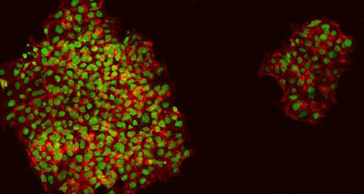
International teams of researchers using advanced gene sequencing technology have uncovered a single genetic mutation responsible for a rare brain disorder that may have stricken families in Turkey for some 400 years.

The discovery of this genetic disorder, reported in two papers in the journal Cell, demonstrates the growing power of new tools to uncover the causes of diseases that previously stumped doctors.

Besides bringing relief to affected families, who can now go through prenatal genetic testing in order to have children without the disorder, the discovery helps lend insight into more common neurodegenerative disorders, such as ALS, also known as Lou Gehrig’s disease, the researchers said.

The reports come from two independent teams of scientists, one led by researchers at Baylor College of Medicine and the Austrian Academy of Sciences, and the other by Yale University, the University of California, San Diego, and the Academic Medical Center in the Netherlands.

Both focused on families in Eastern Turkey where marriage between close relatives, such as first cousins, is common. Geneticists call these consanguineous marriages.

In this population, the researchers focused specifically on families whose children had unexplained neurological disorders that likely resulted from genetic defects.

Both teams identified a new neurological disorder arising from a single genetic variant called CLP1. Children born with this disorder inherit two defective copies of this gene, which plays a critical role in the health of nerve cells.

Babies with the disorder have small and malformed brains, they develop progressive muscle weakness, they do not speak and they are increasingly prone to seizures.

Dr Ender Karaca, a post-doctoral associate in the department of molecular and human genetics at Baylor, first encountered the disorder in 2006 and 2007 during his residency training as a clinical geneticist in Turkey.

“We followed them for years,” said Karaca, a lead author on one of the papers. Karaca said he and his colleagues performed some basic genetic tests on the families but to no avail.

He presented these cases at a genetics conference in Istanbul in 2010, where he caught the attention of Dr James Lupski, who leads the Center for Mendelian Genomics at Baylor, a joint program with the Johns Hopkins University School of Medicine funded by the National Human Genome Research Institute

The center is focused on finding and recruiting thousands of patients and families with undiagnosed disease likely caused by single-gene defects known as Mendelian disorders.

Lupski recruited Karaca into his program at Baylor, where the team continued to work on identifying more cases. A broad genetic test known as exome sequencing, which looks at all of the protein-making genes representing about 1 percent of the genetic code, eventually identified five families with similar characteristics and the same CLP1 mutation.

Researchers needed confirmation in lab tests that the defect could cause the neurological problems seen in the families. That came at a meeting in Vienna, when Dr Richard Gibbs, director of Baylor’s Human Genome Sequencing Center, presented the gene as part of a list of suspected disease-causing variants.

In the audience was Dr Josef Penninger of the Austrian Academy of Sciences in Vienna, whose lab had been working on a model of this same genetic variant in mice.

Both the mice and the people with the genetic defect shared similar characteristics. Further experiments by the Vienna team showed the mutant copies of the CLP1 gene affected the survival of key cells in the brain stem of the mice.

“We had patients with an interesting phenotype (symptoms) and a novel gene but no evidence from the lab that these mutations are disease-causing. They had a model organism, a mouse, but they didn’t have evidence that it affected people. It was a perfect storm,” said Baylor geneticist Dr Wojciech Wiszniewski, author of another study.

As the Baylor-led researchers were working on the problem, collaborators at the Yale Center for Mendelian Genomics, another of the government-funded Centers for Mendelian Genomics, were sequencing families in Turkey under the direction of Dr Murat Gunel, a professor of genetics and neurobiology at Yale.

Gunel and colleague Dr Joseph Gleeson at University of California, San Diego, have been focusing on understanding the fundamental mechanisms of how the human brain develops.

Gunel had been collecting DNA samples from children affected with neurodevelopmental brain problems. The team did exome sequencing on more than 2,000 samples, and they, too, turned up a disorder linked with the CLP1 gene.

Further investigation of the genetic data suggested that all of the cases they identified among four, unrelated families were linked with a single, spontaneous mutation in the CLP1 gene that occurred 16 generations, or about 400 years ago.

Gunel, who received his medical degree in Istanbul, said the high rates of marriages between closely related people in Turkey and the Middle East lead to these rare disorders as affected children inherit mutations in the same gene from both of their parents. Without such marriages, children are very unlikely to inherit two mutations in the same gene.

The Yale paper credits Lupski at Baylor for sharing some of his unpublished findings. Gibbs said the whole effort is an “good example of communication-driven discovery.”

Source: Reuters


Human skin grown in lab ‘can replace animal testing’

hman skin

Skin grown in the laboratory can replace animals in drug and cosmetics testing, UK scientists say. A team led by King’s College London has grown a layer of human skin from stem cells – the master cells of the body.

Stem cells have been turned into skin before, but the researchers say this is more like real skin as it has a permeable barrier. It offers a cost-effective alternative to testing drugs and cosmetics on animals, they say.

The outermost layer of human skin, known as the epidermis, provides a protective barrier that stops moisture escaping and microbes entering. Scientists have been able to grow epidermis from human skin cells removed by biopsy for several years, but the latest research goes a step further.

The research used reprogrammed skin cells – which offer a way to produce an unlimited supply of the main type of skin cell found in the epidermis. They also grew the skin cells in a low humidity environment, which gave them a barrier similar to that of true skin.

Skin barrier
Lead researcher Dr Dusko Ilic, of King’s College London, told BBC News: “This is a new and suitable model that can be used for testing new drugs and cosmetics and can replace animal models.

“It is cheap, it is easy to scale up and it is reproducible.” He said the same method could be used to test new treatments for skin diseases.

Researcher Dr Theodora Mauro said it would help the study of skin conditions such as ichthyosis – dry, flaky skin – or eczema. “We can use this model to study how the skin barrier develops normally, how the barrier is impaired in different diseases and how we can stimulate its repair and recovery,” she said.

The Humane Society International, which works to protect animals, including those in laboratories, welcomed the research, published in the journal Stem Cell Reports.

Research and toxicology director Troy Seidle said: “This new human skin model is superior scientifically to killing rabbits, pigs, rats or other animals for their skin and hoping that research findings will be applicable to people – which they often aren’t, due to species differences in skin permeability, immunology, and other factors.”

Source: BBC news


Thinking Problems Tied to Blockages in Neck Artery

Carotid-Artery-Blockage

Blockage of the carotid artery in the neck appears to increase the odds for memory and thinking problems, a new study indicates.

The researchers said their findings suggest more aggressive treatment might be needed for people with this condition, which is caused by plaque buildup in the artery.

The study is to be presented Wednesday at the American Academy of Neurology (AAN) annual meeting in Philadelphia.

“To date, the focus of diagnosis and management of carotid artery blockages has been prevention of stroke since that was the only harm that these blockages were thought to cause to patients,” Dr. Brajesh Lal, of the Baltimore VA Medical Center and the University of Maryland School of Medicine, said in an AAN news release.

“These results underscore the importance of assessing the status of memory and thinking in people with carotid artery narrowing,” Lal added.

In conducting the study, the researchers examined 67 people with symptomless carotid narrowing, or stenosis. For these participants, the diameter of their artery was cut in half. The study also included 60 people who did not have carotid blockage but did have risk factors, such as diabetes, high blood pressure, high blood cholesterol and coronary artery disease.

Researchers tested participants’ thinking ability, examining their processing speed, learning, memory, decision-making and language.

They found participants with carotid blockage performed much worse on the thinking tests. They also scored lower on motor and processing speed evaluations as well as learning and memory tests. The researchers said language scores did not differ between the two groups of participants.

“If these findings are confirmed in larger studies, they hold significant implications for new treatment targets and open the door for more questions such as: should these patients be treated more aggressively with medications, cognitive rehabilitation, or even surgery to open up the artery,” Lal said in the association news release.

He said he anticipates follow-up studies searching for causes and the best treatment options.

Source: webmd


Researchers find thousands of bacteria living on cash

Man-Counting-Money-Cash-Dollars

The cash in your pocket may be contaminated with so much bacteria that it could make you sick.

Researchers from the Wright Patterson Medical Center in Dayton, Ohio, asked people standing in line at a grocery store checkout and at a high school concession stand to trade a $1 bill from their pocket for a new one. Then the doctors analyzed 68 of those old, worn bills.

Five of the bills contained bacteria that can cause an infection in perfectly healthy people, and 59 of them (that’s 87 percent) were contaminated with bacteria that could cause an infection in anyone with a compromised immune system, such as people with HIV or cancer.

Only four of the bills were relatively clean.

“One-dollar bills are widely used and each is exchanged many times,” said Dr. Peter Ender, one of the study’s authors. “If some are contaminated with bacteria, there is potential to spread these organisms from person to person.”

More Study Needed

So when you hand over the cash, are you giving more of yourself than you intend to? Not necessarily. The study only addresses how much and what kinds of bacteria live on paper money. Another study would be needed to determine whether the money can actually spread the bacteria.

Plus, since the authors only tested 68 bills — there are billions in circulation around the country — the study doesn’t go far enough to prove that as money moves across the country, so do diseases.

Ender says, though, the study does show that paper money is usually full of bacteria, and that a dollar bill could, theoretically, be the magic carpet it rides from one host to another.

But if, in fact, the bacteria can spread on paper money, there’s nothing you can really do about it. You can try to keep your hands away from your eyes, nose and mouth — where the bacteria would get into your body — and try to wash your hands often.

Source: abc news


Infant hair reveals life inside the womb

Hairs can reveal a lot, from your personality to even drug abuse or hormonal changes. Now, add foetus growth in the womb to the hair list.

In a thrilling discovery, a team of researchers including an Indian-origin scientist have found that hair can also reveal the womb environment in which an infant was formed.

They used infant hair to examine the hormonal environment to which the foetus was exposed during development – promising to unleash a wealth of new information in the fields of neonatology, psychology social science to neurology.

“We had this ‘Aha!’ realisation that we could use hair in newborns, because it starts growing one to two months before birth,” said Christopher Coe, director of the Harlow centre for biological psychology at University of Wisconsin-Madison.

“The findings raise questions about everything from the significance of birth order to stereotypical ‘boy’ and ‘girl’ behaviours in children,” Amita Kapoor, an assistant researcher at the Wisconsin National Primate Research Centre, noted.

Additionally, what happens to a developing foetus while in the womb may impact its risk for chronic disease later in life, Kapoor added.

According to researchers, hair closest to the scalp reveals the most recent information but moving down the shaft effectively transits an individual’s hormonal timeline.

For the study, researchers took small samples of hair from mother rhesus monkeys and their infants using common hair clippers. The hair was cleaned and pulverised into a fine powder using a high-speed grinder.

The hormonal signature was then read using a new mass spectrometry method. They found that cortisone, an inactive form of stress hormone cortisol, was higher in young mothers and in their babies than in hair of the older mothers and their infants.

Babies born to young mothers also had higher levels of estrone (a form of estrogen) and testosterone in their hair than did babies born to older mothers.

“Type 2 diabetes, metabolic disease, coronary artery disease and psychiatric disorders – there may be a whole host of long-term
repercussions of stress in utero,” Kapoor emphasised.  The study appeared in the journal Pediatric Research.
Source: business standard


Low blood sugar tied to ‘hangry’ fights with spouse

We’ve all been “hangry,” so hungry that we become angry at the slightest frustration or provocation. But could low blood sugar make you so hangry you’d abuse your spouse?

In an effort find out, scientists asked married couples to secretly stick pins into a voodoo doll representing their spouses, and blast noise in their spouses’ ears. The results, released Monday in the Proceedings of the National Academy of Sciences (PNAS), does appear to show a link between lower blood sugar and marital spats.

Led by Brad Bushman, a professor of communication and psychology at The Ohio State University, the experiment tested a hypothesis about self-control.

The researchers had 107 couples monitor their blood glucose levels with over-the-counter monitors once in the morning and once in the evening for 21 days. Every evening the partners were to privately stick needles into voodoo dolls to indicate how angry they were with their spouses, zero meaning not at all, up to a high of 51.

Even after controlling for a number of variables like overall relationship satisfaction, people with lower glucose levels stuck more pins in the dolls. There was no difference between men and women in how they were affected.

Then Bushman had couples come into his lab to play a simple computer game against each other while sitting in different rooms. In fact, they were playing against a computer and the results were rigged so they’d win and lose about the same number of rounds.

As a punishment for “losing” a round, the victor could play an obnoxious noise — a combination of fingernails on chalkboards and other irritating sounds like an airhorn — into the earphones of the loser at a volume the victor selected, up to about the level of a smoke alarm. (Actually, the computer controlled the noise level.)

Those people with lower glucose levels, and who stuck more pins into the dolls, also tended to blast the noise.

“Thus,” Bushman and colleagues wrote, “low glucose levels might be one factor that contributes to intimate partner violence.”

Many experts believe that self-control can be depleted like a battery, as illustrated in one famous 1998 study: Two groups of hungry people were placed in a room containing a plate of freshly-baked cookies and a plate of radishes. One group was told they could eat cookies, the other told they could eat only radishes. Both groups were then asked to complete a puzzle they didn’t know was unsolvable.

The cookie group worked twice as long on the puzzle. People in the radish group gave up sooner because they had to exercise more self-control to avoid eating the cookies. So there was less willpower left to work on a frustrating puzzle.

A number of factors can deplete self-control, said Brandon Schmeichel, a professor of psychology at Texas A&M University who studies this phenomenon. Performing a task that requires self-control — like not eating a cookie when you really want one, or doing math problems, or filling out your 1040 form — can do it, as can mood, alcohol, and one’s ability to keep your eye on long-term goals, rather than short-term impulses. “That can be difficult to do,” Schmeichel said.

Glucose is a more controversial factor. Proponents argue that the brain uses a lot of energy, especially the pre-frontal cortex that exerts control over our baser instincts and helps us reason. Low glucose can leave the brain low on gas. And being low on gas weakens self-control.

But others point out that some studies suggest self-control is not always limited, and that experiments trying to link low glucose to low self-control are contradictory. Some show an effect, some do not.

Bushman believes there is a cause-effect link and that “aggression starts when self-control stops.”

Professor Florian Lange, a neuroscientist at Hannover Medical School in Germany, praised some parts of the study, but via email said he’s not convinced there’s “a significant role for glucose in self-regulation/self-control.”

A number of other factors could explain the experiment’s results “equally well,” Lange said. For example, he speculated, “who are these violent people having low blood sugar?” he asked.

“Maybe they eat healthier in order to be fit to do extreme sports, an activity they like to pursue because they are more risk-taking,” Lange suggested. “This latter variable could explain why they show more aggression.”

Whether or not low glucose specifically depletes self-control, though, most experts agree that hunger can. As Bushman said, “hungry people are cranky people.”

So, he said, “if you are having a discussion with your spouse about a conflict situation, make sure you’re not hungry.” He advised skipping candy bars and other high-sugar foods, which can spike glucose but lead to a crash. Instead, say, before that last minute tax return debate, eat something nutritious.

source; today


Degenerated organ fully restored in living animal

Scientists have made a breakthrough in regenerative medicine by fully restoring a degenerated organ in a living animal for the first time.

A team from the Medical Research Council (MRC) Centre for Regenerative Medicine, at the University of Edinburgh in the UK, reconstructed the thymus of aging mice, reported medicalnewstoday.com

The thymus is a glandular structure that functions as part of the body’s immune system by creating T cells – the type of white blood cell that is essential for fighting infection.

Located in the front of the heart, the thymus is the first organ to deteriorate as we age.

Scientists have attempted to regenerate the thymus before, using sex hormones. But using this technique, the thymus only temporarily regenerated with limited functional recovery.

In the new experiment, however, the restored thymus was fully functioning and “very similar” to the thymus of a young mouse, say the researchers.

Although the researchers have not yet ascertained whether the immune systems of the mice with a restored thymus were strengthened by the process, they do know that mice receiving this treatment began to produce more T cells.

“One of the key goals in regenerative medicine is harnessing the body’s own repair mechanisms and manipulating these in a controlled way to treat disease. This interesting study suggests that organ regeneration in a mammal can be directed by manipulation of a single protein, which is likely to have broad implications for other areas of regenerative biology,” says a senior MRC researcher.

Source: Press TV


Too much computer time hits bone health in adolescents

Researchers have found that in boys, higher screen time was adversely associated to bone mineral density (BMD) at all sites even when adjusted for specific lifestyle factors.

Results of a study showed that the skeleton grows continually from birth to the end of the teenage years, reaching peak bone mass – maximum strength and size- in early adulthood. Along with nutritional factors, physical activity can also greatly impact on this process.

The Norwegian study explored the hypothesis that greater computer use at weekends is associated with lower BMD. The data was obtained from 463 girls and 484 boys aged 15-18 years in the Tromso region of Norway. The students participated in the Fit Futures study from 2010-2011 which assessed more than 90 per cent of all first year high school students in the region.

BMD at total hip, femoral neck and total body was measured by DXA (dual-energy X-ray absorptiometry). Lifestyle variables were collected by self-administered questionnaires and interviews, including questions on time per day during weekends spent in front of the television or computer, and time spent on leisure time physical activities. The associations between BMD and screen time were analyzed in a multiple regression model that included adjustment for age, sexual maturation, BMI, leisure time physical activity, smoking, alcohol, cod liver oil and carbonated drink consumption.

Source: Business standard


Scientists visualize new treatments for retinal blindness

A new report published in The FASEB Journal may lead the way toward new treatments or a cure for a common cause of blindness (proliferative retinopathies). Specifically, scientists have discovered that the body’s innate immune system does more than help ward off external pathogens. It also helps remove sight-robbing abnormal blood vessels, while leaving healthy cells and tissue intact. This discovery is significant as the retina is part of the central nervous system and its cells cannot be replaced once lost. Identifying ways to leverage the innate immune system to “clean out” abnormal blood vessels in the retina may lead to treatments that could prevent or delay blindness, or restore sight.

“Our findings begin to identify a new role of the innate immune system by which endogenous mediators selectively target the pathologic retinal vasculature for removal,” said Kip M Connor, a researcher involved in the work from the Department of Ophthalmology at the Harvard Medical School and Massachusetts Eye and Ear Infirmary Angiogenesis Laboratory. “It is our hope that future studies will allow us to develop specific therapeutics that harnesses this knowledge resulting in a greater visual outcome and quality of life for patients suffering from diabetic retinopathy or retinopathy of prematurity.”

To make this discovery, Connor and colleagues compared two groups of mice, a genetically modified group which lacked activity in the innate immune complement system, and a normal group with a fully functional innate immune system. Researchers placed both groups in an environment that induced irregular blood vessel growth in the eye, mimicking what happens in many human ocular diseases. The mice that were lacking a functional innate immune system developed significantly more irregular blood vessels than the normal mice, indicating that the complement system is a major regulator of abnormal blood vessel growth within the eye. Importantly, in the normal mice, scientists were able to visualize the immune system targeting and killing only the irregular blood vessels while leaving healthy cells unharmed.

“Knowing how the complement system works to keep our retinas clean is an important first-step toward new treatments that could mimic this activity,” said Gerald Weissmann, Editor-in-Chief of The FASEB Journal. “It’s a new understanding of how proliferative retinopathies rob us of sight, and promises to let us see the path ahead clearly.”

Source: India medical Times