‘Instagram for doctors’ to be launched in Europe

An app which enables healthcare professionals to share photos is to be rolled out across western Europe by the end of the year. The app was designed to enable doctors to share pictures of their patients, both with each other and with medical students.

So far, more than 150,000 doctors have uploaded case photos with the patient’s identity obscured. 'Instagram for doctors' to be launched in Europe
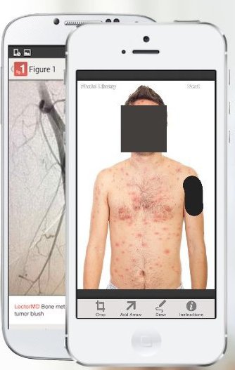
However, some experts have expressed concern about patient confidentiality. Patients’ faces are automatically obscured by the app but users must manually block identifying marks like tattoos.

Each photo is reviewed by moderators before it is added to the database.

No secrets

Founder Dr Josh Landy told the BBC that the Figure 1 service did not access any patient records. “We do not possess any personal medical data at all. The best way to keep a secret is not to have it. We are not an organisation that delivers healthcare,” he told .

But doctors must provide identifying credentials and are also advised to notify their employees and patients to find out about consent policies. “Legally, we found that identifying the doctor does not identify the patient,” said Dr Landy.

“However some [medical] conditions are so rare that they can’t be posted. One user wanted to post something but there are only seven cases of it in the US and they had all been reportable because they are rare, so the patient could have been identified.” Anybody can download the app for free, but only verified healthcare professionals can upload photos or comment on them, he added.

‘Colourful’
“We reject sensationalistic images,” said Dr Landy. “Everything is there for educational purposes. That said, there are very colourful images – things medics see every day. It’s a transparent view into a world you rarely get to see.” The app is already available in North America, the UK and Ireland.

While digital services such as UpToDate and DynaMed – both requiring a subscription – are already widely used within the healthcare community as clinical knowledge databases, they are not rivals to Figure 1, said Dr Landy. “UpToDate is an app I love, and have used for years. However, they have a highly curated repository of articles written and edited by experts in the field.

“What our app does is provide the opportunity to contribute any case no matter how classic or unusual. Ours is all image-based and totally crowdsourced.” The app has received $6m (£3.75m) in investment in the last year. British GP and author Dr Ellie Cannon gave it a cautious welcome.

“I think it’s potentially really useful to share photos with medical students and other doctors,” she said. “Obviously the potential pitfall is the confidentiality. Of course, they are anonymised but even uploading from a certain doctor may go some way to identify a patient,” she added.

“And can a patient later opt out? We’ve seen with other sites the downsides of sharing too much.”

Source: bbc news


Thanks to Google glass: Houston boy virtually ‘visits’ zoo from his hospital bed

Six-year-old Jayden Neal got a glimpse of the Houston Zoo last week, thanks to Google Glass.

Neal has been a regular patient of Children’s Memorial Hermann Hospital in Houston since being diagnosed with type 1 diabetes about a year ago. The hospital recently teamed up with the Houston Zoo and the Google Glass Explorer program to give its pediatric patients a unique experience.

“We had a great time working on the project,” Natalie Camarata, social and multimedia manager at Memorial Hermann, said. “[The hospital] is right across from the Houston Zoo, so it was just a natural fit to be able to partner with them through Google Glass to be able to create that firsthand experience for these kids.”

Neal, who usually looks for animals at the zoo through his hospital room window, was able to get a close-up view of jaguars, sea lions and giraffes by talking with zoo workers through Google Glass..

Camarata says Neal took to the technology very quickly.

“When we were working with him, we told him, you know, you can Google whatever you want. And the first thing he did was Google diabetes,” Camarata said. “That there told me that he not only knew how to use the Internet already, he knew how to use it in a personal way and he was able to navigate this device like nothing.”

The hospital has one pair of Glass through the Google Glass Explorer program. Several pediatrics patients were chosen to participate in the zoo experiment. Though there are no formal plans for any more Glass experiments, Camarata said the reaction has been overwhelmingly positive.

“We have a new appreciation for the device and we’re excited to share this experience with our patients,” Camarata said.

Source: fox news


How Google Glass could revolutionize medicine

web_GoogleGlass_RalfCheung

Here has recently been a slew of media attention to the possibility of an Apple Watch, and the soon to be released Google Glass. For those not familiar with the Google Glass Project, it is essentially a wearable device that mimics eyeglasses.

The hardware includes Wifi and Bluetooth capabilities (can work with Android and iOS devices), cameras, voice-activation commands, and a heads-up display.

This device will find many uses with the general populace, but what about the healthcare field, what does it mean for medicine?

Imagine several medical scenarios using Google Glass:

  • An emergency responder arriving at a motor vehicle accident is able to live stream to the emergency department the status of the patients and the associated trauma suffered to a patient. The ER is then able to assemble and prepare for a patient’s emergency treatment.
  • A surgeon live streams to residents and students a live surgery–so that they can see what work goes into a medical procedure first hand.
  • A visiting nurse seeing a patient in their own home video records and captures images of the patient’s wound (for which they are caring for) and sends them back to the physician.
  • A resident’s physical exam of a patient is streamed back to an attending physician, who can critique their work and make recommendations on questions to ask in real time. This could especially be useful when a resident consultant evaluates a patient while their attending is at home overnight.
  • A cardiologist in a cath lab overlays the fluoroscopy as they perform a femoral catheterization for a patient with a recent myocardial infarct.
  • A nurse scans the medication they are about to give the patient and confirms the correct drug and right patient by overlaying their patient profile with the person in front of them–possibly stopping a medical error.
  • A student brings up their notes and lab reports as they present their patient case to their attending, with data available in real time.
  • An oncologist can overlay the MRI scan over a patient, and show the patient and their family where the cancer exists.
  • The electronic health record at the hospital is available to caregivers, and able to be updated on major changes in the patients they oversee. For instance, the recent cultures from a septic patient’s wound comes back positive for MRSA and the physician changes their broad spectrum antibiotics to appropriate therapy based upon sensitivities.
  • A pharmacist is able to scan medications and verify the proper drugs after comparing the drug with images available in the database, ensuring the right drug is dispensed.
  • A physical therapist can see past sessions with a patient from previous recordings, overlaying their current range of motion, identifying changes as well as progression.
  • Any healthcare professional could walk up to a patient’s bed and instantly see all their vitals such as pulse, BP, O2 Sats, etc.

Could these be major changes that can be implemented by Google Glass or wearable computers? Let’s face it, medicine is changing. We are heavily involved with real time data to treat patients whose status frequently changes. The ability to utilize tools that can keep us connected and up-to-date may help prevent medical errors. It may also increase efficiency of care, collaboration with fellow providers, help educate new students, and lead to a potential major change in medical practice. No longer do we use the black bag of the 19th century physician, but rather we have graduated to using technology to increase our level of care.

Source: Imedical apps


Delhi gets Asia’s first ‘Silent’ MRI system

With a vision of laying as much importance on ‘patient-comfort’ as on the actual medical services provided to them, Mahajan Imaging, north India’s leading chain of high-end medical imaging centres, on Tuesday claimed to have installed Asia’s first ‘Silent’ MRI system in a ‘unique, non-intimidating and non-medical environment’ at its new diagnostic centre in Defence Colony here.

According to Mahajan Imaging, the MR750w MRI system by GE Healthcare has the world’s most advanced imaging technology that enables radiologists to perform MRI scans “without any sound”.

According to GE Healthcare, its revolutionary Silent Scan technology is designed to reduce MR scanner noise to near ambient (background) sound levels and thus improve a patient’s MR exam experience. Conventional MR scanners can generate noise in excess of 110 dBA (decibels) levels, roughly equivalent to rock concerts.

Dr Harsh Mahajan, chief radiologist at Mahajan Imaging, said, “We have installed Asia’s first truly silent MRI system. Till now, MRI scans were associated with a lot of noise – almost as much as a jet engine or rock concert, which led to stress and discomfort for patients. Today, we are able to do most brain scans without any noise at all! This, in addition to the obvious benefit of comfort to the patient, also enables us to do higher quality scans since patients do not move and are more cooperative during scans. This eventually leads to better images and hence a better diagnosis.”

Dr Mahajan said that soon the ‘silent’ technology would be extrapolated to other parts of the body, which would lead to a paradigm shift in MRI scanning.

“The development of this revolutionary Silent Scan MRI Technology is in keeping with GE’s mission of humanizing its medical technology,” said Karthik Kuppusamy, senior director of MR Imaging at GE Healthcare South Asia.

“This machine is also testament of India’s product development capabilities since it was co-developed by our engineers in Bangalore. We used proprietary high-fidelity gradient and ultra-fast RF system electronics and eliminated the sound at the source,” he added.

“It is amazing that there is no drop in image quality,” said Dr Raju Sharma, professor of radiology at the All India Institute of Medical Sciences (AIIMS), New Delhi. “I congratulate GE for developing a clinical system that can perform MRI scans without any sound – this is a dream come true for radiologists all over the world.”

Dr Sharma explained that having a noise-free environment makes the patients very comfortable. This improves utilisation of the MRI machine since scans do not need to be interrupted or repeated frequently.

Additionally, Mahajan Imaging has taken patient comfort a step further by giving both, their MRI room and CT room very unique and aesthetically pleasing interiors. While the MRI room bears the theme — Genesis-Hope-Healing, depicted using a 33 feet long painting of three Greek Goddesses, the CT scan room gives the impression of being underwater.

“The idea is to create an environment of healing,” said Ritu Mahajan, executive director of Mahajan Imaging and the person responsible for designing the facility. “We decided to push the boundaries of what is possible in an MRI room and used silk on the walls and ceiling to add warmth, and matching self-levelling epoxy flooring. Additionally, an artist from Albania was approached to make a 33 feet long painting which depicts Genesis, Hope and Healing, according to Greek mythology.”

“The room housing the CT scan machine in the diagnostic centre has been hand-painted by a young artist to give the impression of being underwater, something that is very popular with younger patients,” she said.

“Since an MRI machine has a very strong magnetic field, and is affected even by extremely small changes in radiofrequency waves, we decided to go with several small specially manufactured LED lights to light up the MRI room,” said Dominique Taffin, senior design architect from GE Healthcare who helped execute the MRI room interiors at the centre.

“The positive effect of one’s surroundings on one’s health cannot be underplayed,” said Dr S S Kale, professor of neurosurgery at AIIMS, New Delhi. “What Dr Mahajan and his team have done is truly unique – every room has a different theme! The environment at the centre sets the patient at ease – a very important component of providing healthcare from a holistic point of view.”

Niharika Dutt, a 22-year old healthcare professional, has had several MRI scans done earlier because of some illness, but feels that this machine is different. “I have had MRI scans done many times because of repeated dizziness and migraine,” said Dutt, “but I have never seen a room this pretty! I had seen photographs of this room online and felt that they were edited – but I later realised that Mahajan Imaging has actually made their room like this. The ambience, in addition to the quietness of the machine, is definitely going to make any patient forget about their disease for some time and make the harrowing experience of the MRI better.”

In addition to the advanced MRI and CT technology, the new centre boasts of the world’s best Mammography machine, X-Ray system, ultrasounds, cardiac scanners, dental CT and DEXA machine – providing all radiology services under one roof.

Source : India Medical Times


New method makes stem cells in about 30 minutes, scientists report

In a feat that experts say is a significant advance for regenerative medicine, scientists have discovered a surprisingly simple method for creating personalized stem cells that doesn’t involve human embryos or tinkering with DNA.

Two studies published Wednesday in the journal Nature describe a novel procedure for “reprogramming” the blood cells of newborn mice by soaking the cells in a mildly acidic solution for 30 minutes. This near-fatal shock caused the cells to become pluripotent, or capable of growing into any type of cell in the body.

When the reprogrammed cells were tagged and injected into a developing mouse, they multiplied and grew into heart, bone, brain and other organs, the scientists found.
“It was really surprising to see that such a remarkable transformation could be triggered simply by stimuli from outside of the cell,” said lead study author Haruko Obokata, a biochemistry researcher at the RIKEN research institute in Japan. “Very surprising.”

The simplicity of the technique, which Obokata and her colleagues dubbed stimulus triggered acquisition of pluripotency, or STAP, caught many experts off-guard.
“So you mistreat cells under the right conditions and they assume a different state of differentiation? It’s remarkable,” said Rudolf Jaenisch, a pioneering stem cell researcher at MIT who was not involved in the study. “Let’s see whether it works in human cells, and there’s no reason why it shouldn’t.”

Obokata said that researchers had already begun experiments on human cells, but offered no details.

Due to their Zelig-like ability to form any number of specialized cells, pluripotent stem cells are considered the basic building blocks of biology. Scientists are working on ways to use them to repair severed spinal cords, replace diseased organs, and treat conditions as varied as diabetes, blindness and muscular dystrophy.

By using stem cells spawned from the patient’s own cells, replacement tissues would stand less of a chance of being attacked by the patient’s own immune system, researchers say. That would spare patients the need to undergo a lifetime regimen of dangerous, immune-suppressing drugs.

But progress toward these lofty goals has been slow, due in part to the challenges of current stem cell production methods. The practice of harvesting stem cells from human embryos makes many people uncomfortable, and some religious groups have pressed for limits or bans on their use. Even scientists who want to study them say they may not be practical for medical therapies because they could be rejected by a patient’s body.

Another approach is to rewind a patient’s own mature cells to a pluripotent state. Dr. Shinya Yamanaka, the first person to make these induced pluripotent stem cells, won a Nobel Prize for this work in 2012. However, the reprogramming process converts only about 1% of the cells into iPS cells, and questions remain about their long-term stability and safety.

The STAP method presents a simpler, cheaper and faster method of producing stem cells, said Chris Mason, a professor of regenerative medicine bioprocessing at University College London.

“How much easier can it possibly get,” Mason told the Science Media Centre, an English organization that promotes scientific understanding on controversial subjects.
“If it works in man, this could be the game changer that ultimately makes a wide range of cell therapies available using the patient’s own cells as starting material,” he said. “The age of personalized medicine would have finally arrived.”

The STAP approach was inspired by observations of plant cells that changed character when they were exposed to environmental stress, according to the research team from RIKEN and Harvard’s Brigham and Women’s Hospital in Boston.

Obokata and her colleagues set about “stressing” mouse blood cells in a variety of ways to see if they would change. They exposed them to heat, deprived them of nutrition and repeatedly poured them through narrow glass pipes.

The method they ultimately published involved placing the cells in an acid solution for 30 minutes and then spinning them in a centrifuge for five minutes. The process converted 7% to 9% of the original cells into STAP cells, Obokata said.

To see whether the cells had been reprogrammed, researchers engineered the mice with a gene that would cause their cells to glow a fluorescent green under ultraviolet light if they became pluripotent. After torturing the blood cells, they began to glow after three days and appeared to peak at seven days, suggesting that they had become pluripotent in just a week’s time. The researchers bolstered the cells’ ability to proliferate by treating them with hormones and an immune cell secretion called leukemia inhibitory factor.

To fully prove that they had become pluripotent, the STAP cells were injected into normal mouse embryos. The resulting offspring, called a chimera, were a mix of regular cells and glowing STAP cells.

Andrew McMahon, director of USC’s Eli and Edyth Broad Center for Regenerative Medicine, said the creation of a chimera was critical to proving that blood cells had changed in a fundamental way.

“That’s the most rigorous [test] you could possibly do,” said McMahon, who was not involved in the study. It shows that the STAP cells can make every type of cell in the embryo and that they “can organize in a normal-looking way, so that what comes out is a normal looking fetus.”

McMahon said the study was also surprising in that it showed that mature cells could be reprogrammed without having to divide.

“That’s why the change is so rapid, because the cells don’t have to undergo division for this to occur,” he said. “It’s a really interesting and novel finding.”

Yamanaka, who was not involved in the STAP study, said the research would undoubtedly help scientists understand the basic biology of cellular reprogramming.

“The findings are important,” said Yamanaka, who directs Kyoto University’s Center for iPS Cell Research.

The reasons why stress causes cells to drastically alter their function remains a mystery, Obokata and her colleagues said.

She declined to say whether the researchers were seeking a patent on the STAP procedure.

Source: latimes