Newborn hearing screening ‘insufficient’ in most provinces

To reach their full potential, children deserve access to proper hearing screening and timely intervention

Too many Canadian babies aren’t being screened for hearing problems at birth, a failure that jeopardizes their future success, say doctors who graded provincial and territorial programs.

Tuesday’s report card from Speech-Language and Audiology Canada and the Canadian Academy of Audiologists finds “serious shortcomings” for newborn screening and comprehensive follow-up.

“The difference between early and late diagnosis of permanent hearing loss on a child’s life is monumental,” Dr. Hema Patel, a pediatrician at Montreal Children’s Hospital and a mother of a son with hearing loss, told a news conference on Parliament Hill.

“As a country, we are failing our children.”

To reach their full potential, all children deserve access to proper hearing screening and timely intervention, Patel said. The ability to communicate effectively with others is the foundation of a child’s social, emotional and educational development.

Extended periods of auditory deprivation can have significant impact on a child’s overall brain development, studies suggest.

Newborns are screened for hearing problems using quick and non-invasive tests that are done by a trained technician, ideally before the infant is discharged from the hospital.

The two hearing groups assigned a grade of “insufficient” to most provinces and territories that offer only localized programs or where the programs were considered to be substandard in quality.

British Columbia was the only jurisdiction to earn a grade of “excellent,” for screening 97 per cent or more of babies across the province with a carefully designed program with clear standards, follow-up and tracking of births and outcomes.

Ontario, Nova Scotia, Prince Edward Island and New Brunswick were considered “good.”

Newfoundland and Labrador, Yukon, Alberta, Quebec, Manitoba, Nunavut, Saskatchewan and Northwest Territories were graded “insufficient.”

The report card is meant to encourage all provinces and territories and the federal government to strive for excellence in early hearing detection programs, Patel said.

Internationally, most universal newborn hearing screening programs recommend screening by one month of age, confirmation of the diagnosis by three months, and intervention by six months, according to the Canadian Paediatric Society’s 2011 position statement.

The report card was endorsed by the Canadian Paediatric Society, VOICE for Hearing Impaired Children and the Elks and Royal Purple of Canada.

Source: CBC


Can Google Glass Transform Medical Education?

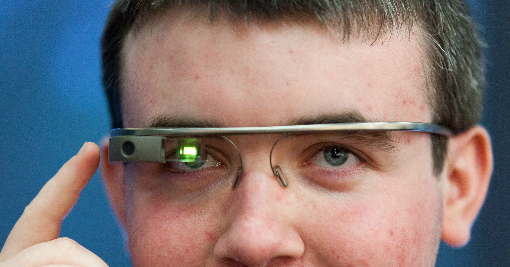
Google Glass looks exciting for the medical world, and presents a particularly powerful opportunity for medical education(for examples, see Forbes article here or Phys.org here). A white paper by the Department of Emergency Medicine, Singapore General Hospital says, “simulation-based training has opened up a new educational application in medicine. It can develop health professionals’ knowledge, skills, and attitudes, whilst protecting patients from unnecessary risks”. Google Glass is taking simulation to the next level and making it more real, as the patients treated are real.

Yet the underlying concept of simulation-based-learning in medicine isn’t new. Neither are the individual components of Google Glass (such as the video recording feature and the possibility of sharing procedures online with any number of students). The biggest innovation might be having all this in one device. As Aristotle said, the whole is more than the sum of its parts.

Medical education is often a two stage process. In stage one, doctors in training need to study voluminous tomes and pass exams; stage one is the collection and storing of knowledge – perhaps too much knowledge. Richard Barker says in his book 2030, the future of medicine, that “as our bio-medical insights continue to fragment traditional diseases into multiple molecular disorders, keeping pace with advances gets tougher and tougher; … ‘head knowledge’ needs to be complemented by online decision support, distilling the wisdom and experience of the best specialist and putting it at the fingertips of the practitioner”. In other words, clinicians are starting to need real-time knowledge on tap.

Stage two focuses on learning through direct patient contact under the guidance of seniors, and Barker’s position suggests that stage two may never really end. Google Glass would support this stage of the curriculum, helping to simulate the practice of medicine, teach decision making, and then allow collaboration long after qualification. With a teacher demonstrating on patients (or that earlier revolution: a mannequin) with a headset camera, the learner is brought straight into the operating theater.

Google Glass is similar to a standard pair of glasses. It has an optical head-mounted display, sitting just above the right eye. Features include a built-in GPS, microphone and Bluetooth, and a camera which can record and live-stream videos to a Google hangout. Particularly useful is voice activation which would allow surgeons to, for example, do a web search for latest research or access EMRs or even real-time patient metrics without “breaking scrub” (compromising operating room sterility). As well as improving the provision of care, this ought to give students a more holistic understanding of each case.

Dr. Rafael J. Grossmann, Surgeon, mHealth Innovator and Google Glass Explorer was the first to perform a Google Glass-aided surgery, including remote teaching contexts and offering clinical advice remotely via Google hang-out. Orthopaedic surgeon Dr. Selene Parekh followed with a demo of foot and ankle surgery, and then plastic surgeon Dr. Anil Shah used the device while carrying out a rhinoplasty. Recently, Medical News Today wrote about a surgeon who live-streamed a procedure using Google Glass and a tablet device.

Grossman says that exposing students to the real life of a surgeon and their problems is critical for training and students should learn and mimic best practices early on. Furthermore, he adds that Google Glass education goes beyond the operating room, “Google Glass is a great start with practically limitless opportunities. “For example, how to connect with patients, how to teach bedside manner, how to prepare patients for surgery can all be best taught from real life examples. Google Glass records it and demonstrates best practice, from A to Z through the responsibilities of a practitioner,” he says.

Plus, of course, these Google Glass recorded procedures can be shared across the globe. Innovator Armando Iandolo, co-founder of Surgery Academy and his team have created an application for Google Glass that lets surgeons stream a heads-up view of procedures to students anywhere in the world. The big, bold innovation is to connect these streams in MOOCs (massive open online courses), says Iandolo. He and his co-founder are currently crowd-funding the idea on Indigogo. “Students will access an operating theatre online and watch a surgical intervention, live, for the procedure of their choice”, says Iandolo. “As we enter Universities, we want to become an integral part of the medical student’s study curriculum”.

MOOCs aren’t new either, but with the Surgery Academy everything seems to fall in place. By bringing the learner straight into theatre, simulation via Google Glass makes courses operate more like apprenticeships.

The patient would need to give their approval, but this is surely quite reassuring for the patient: which practitioner – and one good enough to teach – wants to screw up while being live-streamed to hundreds of students and fellow physicians?

The speed at which Google Glass eventually becomes a standard educational support tool is less certain, and we can learn from previous waves of innovation. In 2010, the Northern Ontario School of Medicine introduced a new mobile device program (medical students received laptops, iPhones and iPads). To assess its value, educators there how medical learners use mobile technologies. Their white paper concluded, “Students would adapt their use of mobile devices to the learning cultures and contexts they find themselves in.” Device value needs to be taught. It depends on how welcome new tech is perceived to be in classrooms, by students, teachers, and the wider ecosystem.

A typical fear is that, especially early in the curriculum (stage one above), medical students will miss out on basic knowledge. Search and find functions make it easier to zero in on an answer, but perhaps without the rich context and basic knowledge provided by reading cover to cover. Students – and teachers – could work just ‘for the test’.

Well, books have always had indices. It’s the process of search which has been accelerated, and there is no evidence that students would treat a digital medical textbook differently than its paperback version. In fact digital isn’t a replacement for the traditional textbook; it’s an opportunity to augment it. There is a generational shift in the learning styles of medical students, Mihir Gupta writes in a KevinMD article. Digital allows the stodgy textbook to be augmented with visual and multimedia, which will suit certain learning styles. “Innovative digital resources are vital for helping students retain knowledge and simplify difficult concepts”, says Gupta. These new resources are great for quick access to updated medical knowledge, but “it will not replace textbook learning, nor should it”.

Lucien Engelen, Director of the Radboud Reshape Center at Radboud University Medical Center, is currently working on various applications for Google Glass in medicine. He says that the only way to get Google Glass into education is “to make it part of education innovation”. He says, “Take some high profile doctors, professors and nurses and some patients and have them run some tests. All of a sudden the advantages (of Google Glass) seem to fall in place seamlessly”.

Frances Dare is Managing Director of Accenture Connected Health Services, which has partnered with Philips on a Google Glass proof of concept. She agrees with Engelen, cautioning that it is important to create an environment in which experimentation can take place and to understand the type of training needed to prepare clinicians to use Google Glass effectively and safely in practice.

But don’t bet against Google Glass. After all, educators have argued for decades over calculators in math class. Engelen says that he really doesn’t think of Google Glass as something special: it’s just another computer form-factor facing the same barriers of acceptance. It will take some time and discussion over privacy to achieve it, but the new wave is coming.

Source: HIT


How Google Glass could revolutionize medicine

web_GoogleGlass_RalfCheung

Here has recently been a slew of media attention to the possibility of an Apple Watch, and the soon to be released Google Glass. For those not familiar with the Google Glass Project, it is essentially a wearable device that mimics eyeglasses.

The hardware includes Wifi and Bluetooth capabilities (can work with Android and iOS devices), cameras, voice-activation commands, and a heads-up display.

This device will find many uses with the general populace, but what about the healthcare field, what does it mean for medicine?

Imagine several medical scenarios using Google Glass:

  • An emergency responder arriving at a motor vehicle accident is able to live stream to the emergency department the status of the patients and the associated trauma suffered to a patient. The ER is then able to assemble and prepare for a patient’s emergency treatment.
  • A surgeon live streams to residents and students a live surgery–so that they can see what work goes into a medical procedure first hand.
  • A visiting nurse seeing a patient in their own home video records and captures images of the patient’s wound (for which they are caring for) and sends them back to the physician.
  • A resident’s physical exam of a patient is streamed back to an attending physician, who can critique their work and make recommendations on questions to ask in real time. This could especially be useful when a resident consultant evaluates a patient while their attending is at home overnight.
  • A cardiologist in a cath lab overlays the fluoroscopy as they perform a femoral catheterization for a patient with a recent myocardial infarct.
  • A nurse scans the medication they are about to give the patient and confirms the correct drug and right patient by overlaying their patient profile with the person in front of them–possibly stopping a medical error.
  • A student brings up their notes and lab reports as they present their patient case to their attending, with data available in real time.
  • An oncologist can overlay the MRI scan over a patient, and show the patient and their family where the cancer exists.
  • The electronic health record at the hospital is available to caregivers, and able to be updated on major changes in the patients they oversee. For instance, the recent cultures from a septic patient’s wound comes back positive for MRSA and the physician changes their broad spectrum antibiotics to appropriate therapy based upon sensitivities.
  • A pharmacist is able to scan medications and verify the proper drugs after comparing the drug with images available in the database, ensuring the right drug is dispensed.
  • A physical therapist can see past sessions with a patient from previous recordings, overlaying their current range of motion, identifying changes as well as progression.
  • Any healthcare professional could walk up to a patient’s bed and instantly see all their vitals such as pulse, BP, O2 Sats, etc.

Could these be major changes that can be implemented by Google Glass or wearable computers? Let’s face it, medicine is changing. We are heavily involved with real time data to treat patients whose status frequently changes. The ability to utilize tools that can keep us connected and up-to-date may help prevent medical errors. It may also increase efficiency of care, collaboration with fellow providers, help educate new students, and lead to a potential major change in medical practice. No longer do we use the black bag of the 19th century physician, but rather we have graduated to using technology to increase our level of care.

Source: Imedical apps


Smartphone gets smarter, can detect eye defects

Apps and downloads are old hat. The smartphone can now be adapted to test a person’s eyes, and is claimed to be India’s first mobile phone ophthalmoscope.

The phone-cum-scope can capture high-quality photographs of the retina in the eye to detect defects. Converting the smartphone to an ophthalmoscope is quick and easy: fix an LED bulb near the camera of the mobile, a connecting wire and battery. It costs not more than Rs 35-50. The device was invented by ophthalmologists of Sankara Eye Hospital. These pictures can then be sent to doctors for diagnosis.

“We aren’t sure if patients can use it themselves. But it helps eye specialists in rural areas who probably don’t have high-end ophthalmoscopes. We are networking with doctors in rural areas who can send us photographs of patients’ eyes taken through a mobile phone, that can help evaluate the disorder. We’ve had cases of eye tumours, glaucoma and other disorders related to the optic nerve, detected through photos taken from mobile phones with a minimum 5-mega pixel camera,” said Dr Divyansh K Mishra, retina fellow, Sankara Eye Hospital, who was one among the doctors who invented the new technique.

A team of ophthalmologists consisting of Dr Mishra, Dr Madhu Kumar, Dr Rajesh R, Dr Srinivasulu Reddy and Dr Gladys Rodregues, led by Dr Mahesh P Shanmugam, is the think-tank behind this innovation.

Back in 2002, Dr Shanmugam had tried taking pictures of eyes through a handycam. “Technology is now advanced and some cell phones have 21 pixels. We won’t stop using ophthalmoscopes but this is an added advantage. Eye care field technicians can now immediately diagnose a serious eye condition as they’re able to instantly send images of the patient’s retina to doctors in the city, via social networking sites,” said Dr Mishra.

MIT’s mobile gadget

In 2010, a team of students from Massachusetts Institute of Technology came up with a device to be attached to a mobile phone that can test the visibility of eyes and find out the refractive errors. It was called NETRA (Near-Eye Tool for Refractive Assessment). The user has to place the device in front of the eye and look into the lens attached to the phone. While pressing the arrow keys, the user can see green and red lines which overlap. The same process is repeated eight times, with the formation of green and red lines on the screen at different angles. The assessment of the eyes is done based on the visual index and prescriptions are made based on the same.

Source: Times of India


Low-cost ultrasound machines may help reduce maternal mortality in rural areas

In one of the US Government’s Department of Health and Human Services’ reports for grants and guides, there has been a mention of the prerequisite of developing handheld ultrasound devices, especially for the detection of abscesses of lymph node enlargement, applicable to patients with HIV positive or negative. However, the need of ultrasound in medical services is huge as it is most popularly used for basic screening of high-risk pregnancies, cardiac, vascular, radiology, endocrinology and paediatric purposes. “It is most cost-effective and non-radiating machine that is used in any hospital across the globe. For a doctor who wants to have a quick insight of the reason behind the stomach pain, ultrasound is an immediate choice of diagnosis,” S Ganesh Prasad, director, ultrasound imaging, GE Healthcare, told India Medical Times.

Cost-Factor

According to Ganesh Prasad, ultrasound devices have existed in India for 20 years now but the significant market growth has happened in the last 10 years. “Prior to this, the ultrasound devices were available everywhere for the cost of 50,000 dollars (Rs 30 lakh) each but with the introduction of medical technology, low-cost devices have been produced and brought to various customers. Today, ultrasound machines are distributed for the cost ranging from Rs 2 lakh to Rs 70 lakh to various hospitals and medical experts.”

He further said, “The ultrasound machine worth Rs 2 lakh is a simple machine with which you can only do a simple scan to see womb or foetus, while the advanced ultrasound technology with higher cost provides you the real time assistance in witnessing the dynamic evolution of a foetus.”

GE Healthcare is doing a lot of research and development to improve the high quality ultrasound machine’s price form. “Our low-cost ultrasound devices range starts from Rs 4 to 5 lakh,” informed Ganesh Prasad.

“The cost of ultrasound machines depends on different gradations that the machine offers. Some are high-end machines of which prices go up to Rs 1 crore, mid-end machines cost around Rs 50 lakh and low-end machines are good enough for all the purposes, which cost up to Rs 12 to 15 lakh,” Dr Rakhee Gogoi, head of radiology, Paras Hospitals, Gurgaon, told India Medical Times while giving an estimate on the cost of the machines.

Quality Vs Low Cost

“We don’t believe in cheaper performance machine. We make products, which are good for the customers in terms of high technology. By the end of the year, we are planning to release a tablet ultrasound for the first time ever in the world. Every occupation could have it. For example, when a pregnant lady goes for a screening or check-ups, most of the time it’s only the physical exam that’s being done. Many a times, at the time of complication during delivery, there is no machine available for the doctors to see and learn the condition of foetus inside. The whole objective behind this technology is to make it relevant for you. We focus on target customer group and capability of technology, bringing it to a point of price that’s affordable,” said Ganesh Prasad.

GE’s low cost ultrasound devices come at the price point of Rs 5 lakh and products with a valid portfolio come with three years warranty. “We deploy current technology, not compromising in the quality at all levels. The whole idea behind offering three years warranty is to focus on quality as we understand that maintenance charges is a very good factor for the sustenance of ultrasound machines to a customer. We give the quality of a high-end machine but at the lower cost for people,” said Manoj Menon, communication leader, GE Healthcare India.

Penetrating Rural India

“Our villages do not have many ultrasound machines in rural areas. Ultrasound is very basic and important modality that can be used to detect any sort of pathology at a very early stage. Often, villagers come for the treatment when it’s too late for the problem till then the condition has gone out of hand. This is the reason why introduction and distribution of low-cost ultrasound machines will be really helpful in the rural areas,” said Dr Gogoi.

GE Healthcare is already planning to introduce low-cost ultrasound machines in the rural areas of India. “We have divided the market in tiers. Tier I is metro, tier II involves cities like Jaipur and tier III, IV and V have rural districts under them. We are doing good in tier I and tier II. In the tier III market, we have just stepped into. Besides producing the equipment, we are also creating a distribution network and affordability packages that has to be taken further,” said Menon.

Ganesh Prasad asserted, “Tier III is a fastest growing market with 22-23 per cent growth and that is going to be our area of focus. The reason why we have not penetrated as much in the tier III is because of the availability of the cost price for this kind of market. Our main effort has been to build a distribution network around tier II and the more challenging is going to be creating distribution around tier III.”

Improving Healthcare Situations

India has a high maternal mortality rate. “By proper screening during pregnancy, we can see improvement in the outcomes and avoid maternal or infant death during delivery. Ultrasound is the only device that can be used on a pregnant lady. Secondly, in rural areas, deliveries are still done at home which is intoxicating for a mother as well as for a child. With the introduction or availability of ultrasound machines in hospitals or nursing homes in tier III areas, pregnant women can be taken to hospitals for screenings. Some states have already taken the lead as Gujarat has improved its IMR rates and is moving towards greater institutional deliveries, while Tamil Nadu has also seen dramatic improvements in the reduction of infant mortality,” said Ganesh Prasad.

For Dr Jitendra Sharma, head, division of healthcare technology, National Health System Resource Centre, Ministry of Health and Family Welfare, the idea of handheld ultrasound devices clinically would be great and cost-effective. “It would be better than static ultrasound which comes for lakhs of rupees. What we are concerned about is social and regulatory aspect. Even with the static ultrasound machines in hospitals, we have not been practically able to control female abortions. If the hand held devices are sold in the market, let that market be rural or urban, how would female foeticide be controlled,” he told India Medical Times.

Controlling the Misuse for Sex Determination

Dr C V Bhirmanandham, vice president, Medical Council of India, told India Medical Times, “Why spend so much money when we can get good results from a low-cost ultrasound machine? But it should not be misused. The medical practitioners using the machines should be registered with a concerned authority and should be regularly monitored. Anyone caught misusing the machines for sex determination should be readily punished and their license should be cancelled.”

Dr Bhirmanandham also emphasised on the certification that the trainee must get from the concerned authority. “They have to follow the government of India policies and have to be ethical in their approach,” he added.

According to the Pre-Conception and Pre-Natal Diagnostic Techniques (PCPNDT) Act, 1994, the law prohibits any person from using ultrasound equipment for the detection of the sex of an unborn child. With an amendment in 2002 to the PCPNDT Act, the manufacturers and distributors of the ultrasound machines have to ensure the proper use of the equipment and are prohibited from selling, renting, permitting or authorizing the use of ultrasound machines for foetal sex determination. Manufacturers of ultrasound machines must also confirm that the customer has a valid PCPNDT certificate and they are also to provide the government with a quarterly report disclosing to whom the equipments have been sold.

“Hence, we don’t sell any of our ultrasound equipments to customers who are not registered under the PNDT Act. However, we believe that limiting the access of technology is more dangerous because we talk of saving lives and with the use of ultrasound technology, lives with different diagnosis can be saved. As per the government policies, we also continue to report to the government in every three months, disclosing about our dealings,” said Ganesh Prasad.

Source; India Medical Times


Doctors Use 3-D Printing To Help A Baby Breathe

Ever since the day Garrett Peterson was born, his parents have had to watch him suddenly just stop breathing.

“He could go from being totally fine to turning blue sometimes — not even kidding — in 30 seconds,” says Garrett’s mother, Natalie Peterson, 25, of Layton, Utah. “It was so fast. It was really scary.”

Garrett was born with a defective windpipe. His condition, known as tracheomalacia, left his trachea so weak the littlest thing makes it collapse, cutting off his ability to breathe.

“When he got upset, or even sometimes just with a diaper change, he would turn completely blue,” his mother says, “and that was terrifying.”So the Petersons contacted Dr. Glenn Green at the University of Michigan, who specializes in conditions like Garrett’s. He teamed up with Scott Hollister, a biomedical engineer who runs the university’s 3-D Printing Lab, to create a remarkable solution to Garrett’s problem — a device that will hold open Garrett’s windpipe until it’s strong enough to work on its own.

Instead of shooting ink onto a flat page to print words or pictures, 3-D printers use other material, such as plastic or metal, to create three-dimensional objects. “You build up layers until you have the complete 3-D structure,” Hollister says.

3-D printers have been used to build jewelry, art and even guns. But Hollister is using the technology to create medical devices. He uses a 3-D printer that melts particles of plastic dust with a laser. He has already built a jawbone for a patient in Italy and has helped another baby with a condition similar to Garrett’s. But Garrett is a lot of sicker and his condition is a lot more complicated.

“It’s just been issue after issue with breathing, and just trying to keep him breathing at all,” Jake Peterson, Garrett’s dad, says.

At 16 months old, Garrett had never been able to leave the hospital. Every time he stopped breathing, it was a mad rush to save him. And the doctors weren’t sure how much longer they could keep him alive.

“In some sense we were thrown directly into the fire,” Hollister says. “We characterized it as sort of a Hail Mary pass.” So they rushed Garrett from Salt Lake City to Ann Arbor on Jan. 18 and got to work.

First they took a CT scan of Garrett’s windpipe so they could make a 3-D replica of it. Next they used the 3-D printer to design and build a “splint.” It’s a small, white flexible tube tailored to fit around the weakest parts of Garrett’s windpipe.

“It’s like a protective shell that goes on the outside of the windpipe and it allows the windpipe to be tacked to the inside of that shell to open it up directly,” Green says.

But the device has not been approved by the Food and Drug Administration. So Green and Hollister had to convince the agency to give them an emergency waiver to try it. And they were running out of time. “His condition was critical. It was urgent and things needed to be done quickly. It was highly questionable whether he would survive and how long he would survive,” Green says.

Garrett’s parents knew they were taking a leap of faith. But they felt like they had to try. “We were just so excited for that glimmer of hope that this could be what would help Garrett get home,” Jake Peterson says.

Hollister and Green got the FDA’s approval and scheduled the surgery for Jan. 31. As soon as the surgeon, Dr. Richard Ohye, opened up Garrett’s chest, he and Green could see that Garrett’s windpipe had collapsed. One of his lungs was completely white.

“The only time I’d seen a white lung was in somebody that had died,” Green says. They quickly got to work, gingerly placing the first of two splints on one side of Garrett’s windpipe. It fit perfectly. So they got started on a second splint, which fit perfectly, too.

After more than eight hours, both splints were securely in place. Then came the most important moment: What would happen when they let air flow through Garrett’s windpipe into his lungs?

This time, Garrett’s windpipe stayed open, and his white lung turned pink. “That was just amazing to me,” Green says. “Here something that we’d worked on, that had been constructed just a week ago to match this defect. It had worked just the way we had hoped. I said, ‘This is going to change this boy’s life and his family’s life forever.’ ”

Garrett is 18 months old now and is still in the hospital, but in the weeks since the surgery, he has gotten stronger and stronger and needs less help breathing. His parents are ecstatic.

“He has been doing so good. He’s been smiling, and it’s crazy to be able to see him get really upset and not change colors,” Natalie Peterson says. “He’s being more interactive and more alert and reaching more for his toys. He’s just starting to be more like a normal child,” Jake Peterson adds. Garrett’s splint is designed to expand as he grows and eventually dissolve in his body as his own windpipe gets strong enough to work normally.

Green wants to save more babies this way, but it’s expensive to transport these extremely sick children across country. It has also been hard to convince insurance companies to pay for the trip.

“It is one of the most frustrating things that I’ve been through, knowing that there’s something that we have that can help and looking at all the roadblocks that are in place,” Green says. So he’s hoping to launch a formal study, which may enable him to try more splints to save more babies.

Green says this is the most exciting thing he has seen since medical school. “We’re talking about taking something like dust and converting it into body parts,” he says. “And we’re able to do things that were never possible before.” They’ve already started using 3-D printing to build more body parts, including ears and noses, by combining the plastic structure with human cells. Other scientists have gone even further, using 3-D printing to make blood vessels, skin and even primitive organs out of cells.

Source: npr


Internet-derived health insights are now routinely sought by patients

People are increasingly turning to the Internet to share their health problems on the Internet, seeking insights and practical advice, an Oxford study has found. Researchers said the last decade has seen a remarkable shift in how people use the Internet in relation to their health, and it is now talked of as a routine feature of being ill.

The study examined patient interviews conducted between 2001 and 2013, and explored how people talked about the Internet, capturing changing attitudes towards the use of the Internet for health across the last decade.

In the early 2000’s, people who sought health information online saw themselves as particularly engaged, expert and activated patients.

By 2013, the web had become an almost routine part of many people’s experience of health and illness, researchers said.

“The Internet has transformed how people make sense of and respond to symptoms, decide whether to consult, make treatment choices, cope with their illness and connect to others,” said Professor Sue Ziebland from the University of Oxford.

The study found that people want more than just information online, they also seek reflections, insights and practical advice from other patients.

“Every year millions of people use sites such as Oxford’s www.healthtalkonline.org to learn about their health issues from other patients,” researchers said.

Film, animations, sound, pictures and personal experiences online make health information more digestible for people from all backgrounds.

“By helping people to learn about their condition, prepare for consultations and demonstrate to doctors their interest and involvement, the web may even help to undermine some health inequalities,” said Ziebland.

Increasingly, doctors are aware of the trend, and recommend useful sites to their patients. However, even in 2013, patients were reluctant to talk to their doctors about what they find online, fearing that such revelations might damage their relationship with their doctor.

“GPs and nurses who recognise that people are using the Internet when they are ill can support and discuss the information with their patients; those who do not recognise this shift can unwittingly undermine and patronise their patients,” Ziebland said.

Source: ndtv


Aquarium pumps saving babies’ lives

When Chokonjetsa was born, he weighed just over 2 pounds. His tiny, underdeveloped lungs struggled for each breath. Then doctors in the hospital in Blantyre, Malawi, put Chokonjetsa on a breathing machine made out of aquarium pumps.

Chokonjetsa is one of dozens of newborns in the African country who have survived thanks to the ingenuity of students in a senior design class at Rice University in Houston. The overall survival rate of babies with respiratory distress at the Malawi hospital increased from 44% to 71% with the use of the students’ breathing machine, according to a recent clinical trial.

Using aquarium pumps, the students have designed a low-cost version of a breathing device called a bubble CPAP, or Continuous Positive Airway Pressure, machine. Traditional bubble CPAP systems used in developed countries can cost upwards of $6,000; the students’ machine can be manufactured for around $350, providing big savings for hospitals in poverty-stricken areas.

Malawi has the highest rate of preterm birth in the world, 18.1 per 100 live births, according to the World Health Organization. Babies born too early often have underdeveloped lungs that don’t inflate easily.

Premature babies struggle to breathe in part because they don’t produce enough lung surfactant, a naturally-produced chemical that keeps the tiny air sacs in the lungs, called alveoli, from collapsing. A lot of these babies die because of the stress struggling to breathe puts on their bodies.

Second baby possibly ‘cured’ of HIV

In Malawi, when a newborn is suffering from respiratory distress, he or she is typically provided extra oxygen via tubes placed gently in the nose. But doctors don’t add air pressure to help inflate the infant’s lungs. This is the way respiratory distress was treated in newborns, even in developed countries like the United States, until the 1970s.

With a CPAP machine, gentle air pressure is pushed via tubes into the nostrils and/or mouth to help facilitate breathing.

To understand how a bubble CPAP works, think of a straw that is partially submerged in a glass of water. When you blow into the top of the straw, bubbles come out of the bottom of the straw and bubble up.

“When lungs are underdeveloped, it’s like when you’re blowing up a completely deflated balloon and that first breath to inflate the balloon is really difficult,” said Dr. Rebecca Richards-Kortum of Rice University, who helped facilitate the design of the students’ bubble CPAP machine. “For a baby with underdeveloped lungs, every breath is like that.”

Doctors and nurses know that it takes very little air pressure to assist newborns. The design students found that two aquarium pumps provided the perfect amount of gentle pressure for delicate lungs. In the original prototype, the machine was housed in a clear plastic shoebox from Target.

The device’s latest casing box is made from bent sheet metal specifically designed for usability, funcation and durability, said Dr. Maria Oden of Rice University, a co-author of the device’s clinical trial along with Richards-Kortum.

“Having used bubble CPAP myself, the (low-cost aquarium pump) machine uses the same general principles,” said Dr. Andrea Trembath, a neonatal specialist at Rainbow Babies and Children’s Hospital in Cleveland who was not associated with the device’s design. “We’ve found bubble CPAP to be very beneficial in infants with respiratory distress. … After taking a look at the photos, the low-cost machine doesn’t seem to be very different.

Jocelyn Brown, a former student who helped design the low-cost bubble CPAP machine, has lived in Malawi for almost two years. She is collaborating with aid organizations and using a transitions grant from USAID in hopes of rolling out the device across the country — first in government hospitals, and then in private and rural hospitals.

So far, the machines have been implemented in nine government hospitals, she said.
Mysterious cluster of birth defects stumps doctors
Brown said one major challenge to the rollout in Malawi is a lack of nurses and physicians; the high turnover rate makes training difficult.

“Many district hospitals have maybe one doctor, who may not even be a neonatal specialist,” Brown said. “Much of the care is provided by nurses, but one nurse could be taking care of 40 patients at a time.”

Another challenge, Brown explained, is that parents in Malawi are sometimes reluctant to allow the bubble CPAP machine to be used, perhaps because nasal tubes and masks used in respiratory therapy are associated with death — usually, only extremely ill patients receive breathing assistance.

Fortunately, baby Chokonjetsa’s family allowed him to receive the respiratory therapy. His name, which means “thrown away” in Malawi’s Chichewa language, was chosen by his grandmother. She feared he had been thrown away by God, as she didn’t expect him to survive.

But with the help of a breathing machine made of aquarium pumps, Chokonjetsa is now a healthy, thriving 7-month-old baby.

Source: CNN news


Pioneering 3D printing reshapes patient’s face in Wales

A survivor of a serious motorbike accident has had pioneering surgery to reconstruct his face using a series of 3D printed parts. Stephen Power, from Cardiff, is thought to be one of the first trauma patients in the world to have 3D printing used at every stage of the procedure.

Doctors at Morriston Hospital, Swansea, had to break his cheekbones again before rebuilding his face. Mr Power said the operation had been “life-changing”. The UK has become one of the world’s pioneers in using 3D technology in surgery, with advances also being made by teams in London and Newcastle.

While printed implants have previously been used to help correct congenital conditions, this operation used custom-printed models, guides, plates and implants to repair impact injuries months after they were sustained.

Despite wearing a crash helmet Mr Power, 29, suffered multiple trauma injuries in the accident in 2012, which left him in hospital for four months. “I broke both cheekbones, top jaw, my nose and fractured my skull,” he said.

“I can’t remember the accident – I remember five minutes before and then waking up in the hospital a few months later.” In order to try to restore the symmetry of his face, the surgical team used CT scans to create and print a symmetrical 3D model of Mr Power’s skull, followed by cutting guides and plates printed to match.

Maxillofacial surgeon Adrian Sugar says the 3D printing took away the guesswork that can be problematic in reconstructive work.

“I think it’s incomparable – the results are in a different league from anything we’ve done before,” he said. “What this does is it allows us to be much more precise. Everybody now is starting to think in this way – guesswork is not good enough.”

The procedure took eight hours to complete, with the team first having to refracture the cheekbones with the cutting guides before remodelling the face.

‘Life changing’
A medical-grade titanium implant, printed in Belgium, was then used to hold the bones in their new shape.

Looking at the results of the surgery, Mr Power says he feels transformed – with his face now much closer in shape to how it was before the accident.

“It is totally life-changing,” he said. “I could see the difference straightaway the day I woke up from the surgery.” Having used a hat and glasses to mask his injuries before the operation, Mr Power has said he already feels more confident.

“I’m hoping I won’t have to disguise myself – I won’t have to hide away,” he said. “I’ll be able to do day-to-day things, go and see people, walk in the street, even go to any public areas.”

The project was the work of the Centre for Applied Reconstructive Technologies in Surgery (Cartis), which is a collaboration between the team in Swansea and scientists at Cardiff Metropolitan University.

Design engineer Sean Peel has said the latest advance should encourage greater use of 3D printing in the NHS. “It tends to be used for individual really complicated cases as it stands, in quite a convoluted, long-winded design process,” he said.

“The next victory will be to get this process and technique used more widely as the costs fall and as the design tools improve.” Mr Power’s operation is currently being featured in an exhibition at the Science Museum in London, called 3D Printing: The Future.

Source: BBC news


U.S. FDA approves first device to prevent migraine headaches

The U.S. Food and Drug Administration (FDA) said Tuesday it has approved the marketing of a medical device as a preventative treatment for migraine headaches.

The FDA said in a statement that this is the first transcutaneous electrical nerve stimulation (TENS) device specifically authorized for use prior to the onset of pain.

The device, to be marketed under the name Cefaly, is manufactured by STX-Med in Herstal, Liege, Belgium.

“Cefaly provides an alternative to medication for migraine prevention,” said Christy Foreman, director of the Office of Device Evaluation at the FDA’s Center for Devices and Radiological Health. “This may help patients who cannot tolerate current migraine medications for preventing migraines or treating attacks. ”

Cefaly is a small, portable, battery-powered, prescription device that resembles a plastic headband worn across the forehead and atop the ears, the FDA said. The user positions the device in the center of the forehead, just above the eyes, using a self- adhesive electrode.

The device applies an electric current to the skin and underlying body tissues to stimulate branches of the trigeminal nerve, which has been associated with migraine headaches. The user may feel a tingling or massaging sensation where the electrode is applied, the agency said.

According to the FDA, Cefaly is indicated for patients 18 years of age and older and should only be used once per day for 20 minutes.

The FDA said the approval was based on data from a clinical trial in Belgium involving 67 individuals who experienced more than two migraine headache attacks a month and who had not taken any medications to prevent migraines for three months prior to using Cefaly.

The study found that those who used Cefaly experienced ” significantly fewer days” with migraines per month and used less migraine attack medication than those who used a placebo device, the agency said.

The approval was also based on a patient satisfaction study of more than 2,300 Cefaly users in Belgium and France, which showed that about 53 percent of patients were satisfied with Cefaly treatment and willing to buy the device for continued use, it said, noting that no serious adverse events occurred during either study.

Source; News