Get dangerous germs out of your home

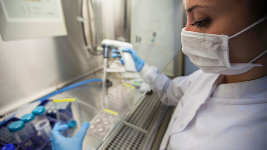
Even if you’re one of the many people who believe that exposing yourself to day-to-day germs is healthy for your immune system, it’s still wise to take steps to protect yourself from the most infectious germs in your home.

“Bugs like Escherichia coli (E.coli), salmonella and campylobacter can make you pretty sick or even kill you,” says Douglas Powell, professor of diagnostic medicine and pathobiology at Kansas State University in Manhattan, Kansas, and author of Barfblog.com. The Centers for Disease Control and Prevention estimates that seven pathogens cause about 90% of these illnesses, hospitalizations and deaths. Scientists dub them “food-borne illnesses” because they’re microbes that invade your body through the gastrointestinal tract.

(Considering that we put our hands to our faces anywhere from 18 to 40 times per hour, is it any surprise that our mouths are the primary point of entry?)

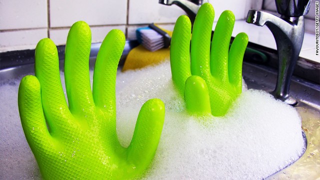
You’ll recognize the poisoning by the fever, nausea, vomiting and diarrhea that results. Luckily, it isn’t too hard to eliminate the dangerous microbes you may unknowingly drag into your home. “Chlorine bleach is your friend,” says Powell, referring to one of the most effective toxin-destroying products around. Rooting out the microbes’ whereabouts, on the other hand, can be trickier. Read on, and you’ll learn how to eliminate from your home the microbes and toxins that could affect your family’s health.

Good: Rearrange your fridge to reduce your risk

You don’t have to travel to Latin America to experience a case of Montezuma’s Revenge. Each year, one in six Americans contracts food poisoning right here on U.S. soil, according to the CDC. In fact, a mere trip to your refrigerator could put you at risk. “The raw meat you bring home from the grocery store has bacteria that can result in diarrheal disease,” says Dr. Rose Devasia, assistant professor at University of Louisville School of Public Health And Information Sciences in Louisville, Kentucky.

A recent report from the Center for Science in the Public Interest listed ground beef, chicken, turkey and steak as the most susceptible to disease. Devasia recommends double-bagging the meats or placing them on the bottom shelf of your refrigerator on a plate, away from the likes of the apples, strawberries or celery you’ll eat cold.

“You’ll go on to cook your meat to kill these microbes,” she says, “but you don’t want that slimy liquid from the package to spill onto the fruits and vegetables you eat raw.”

Better: Place toothbrushes far away from the john

You wouldn’t lick your toilet on purpose. Yet leaving a damp, exposed toothbrush within three feet of the loo isn’t much better. “Studies have shown that bacteria in the toilet can disperse in the air after flushing it,” says Devasia. “Toilet water — along with whatever you’ve deposited in it — gets aerosolized and lands onto what’s nearby, like a toothbrush or hand towel.”

A recent University of Manchester study found that the average toothbrush contained about 10 million germs, including E. coli and staphylococci. An easy fix: Put the lid down on the toilet before you yank the lever to dispose of the contents.

Adds Devasia: “We all know the bathroom is not the cleanest place, so wash your hands to avoid getting yourself sick.” Soaping up at the sink can reduce your chance of getting ill by 30%.

Best: Leave your shoes at the front door

Sleeping with the enemy may only be a closet door away. If you walk through your house with your shoes on, you may be dragging in all kinds of nasty germs and chemicals from the great outdoors. The bottoms of your shoes spread many unhealthy agents, from pollen and pesticides on the lawn to salmonella in bird poop.

In fact, as many as nine different kinds of pathogens can thrive on shoes, according to a University of Arizona study. Microbes can survive and even multiply because of nutrient-rich soil and other deposits left on the soles. “In my house, we take our shoes off when we enter,” says Devasia. “Why drag in a bunch of dirt and dust? Even if you clean floors and carpets regularly, there is some level of dirt that remains.”

Source:  http://www.medicalwebtimes.com/read/get_dangerous_germs_out_of_your_home/


Man lives without breathing for a month

A year ago, 42-year-old runner Victor Carlos never thought he’d be excited just to see the starting line. But that was before he almost died.

In fact, the father of two girls didn’t breathe for a month; a machine pushed oxygen through his body.

Now he’s training for another marathon.

It all began in December with a bad bout of the flu that led to an acute bacterial infection. By the time he checked into the hospital, Carlos was barely hanging on.

“We walked into the triage, and his oxygen level was only 57%, and everybody turned a corner,” Carlos’ wife, Brenda Voglewede, told CNN. Healthy blood oxygen levels are higher than 95%.

Carlos was in deep trouble. He had developed acute respiratory distress syndrome, also known as ARDS. It was slowly killing Carlos by attacking his lungs.

“Not only that, but he had multiple organ failure related to the infection. So his kidneys were not working. His liver was not working. And his bone marrow was failing as well,” said Dr. Ashok Babu, a cardiothoracic surgeon at the University of Colorado Hospital.

“The lungs basically collapse down to nothing. … They are just resting.”

Dr. Ashok Babu

Babu had no time to spare. He knew trying to force air into Carlos’ lungs would do more harm than good. But without oxygen, Carlos’ organs would soon shut down. Babu had one shot: a procedure called extracorporeal membrane oxygenation, or ECMO.

With this treatment, doctors turn down the patient’s respirator, then insert a special tube through the heart. A pump draws blood from the body, flushes it through an oxygenation machine that removes the carbon dioxide and delivers oxygen back to the heart. It functions as an external lung, without the patient taking a single breath.

“So (oxygenated blood) could pass through the lungs, and the lungs didn’t have to do anything to it because it was already processed,” Babu said. “And what we see on the X-ray is the lungs basically collapse down to nothing. … They are just resting. We support the patient until the lungs can heal on their own.”

Carlos’ family was told he had a 40% to 50% chance of survival.

“Dr. Babu initially said he thought he’d be on ECMO for a week,” Voglewede recalled. “Then the week turned into two weeks. And then he’s just like, ‘Well, sometimes you just have to wait these things out.’ ”

Babu said, “Two weeks into it we were all pretty worried that we had this young guy whose lungs just didn’t seem to be coming back. At the time, he was the patient we had had on ECMO the longest in the history of our program.”

Two weeks became four. Carlos didn’t take a single breath for a month.

Then a breakthrough came.

“Somewhat miraculously, his lungs just started to open up on their own,” Babu said.

By that time, Carlos was a shadow of his former self. He had lost more than 30 pounds.

“You can imagine, before going into the hospital you are this active person. Then you go in, they put you under, and when you wake … you can’t sit up, you can’t move your arms up and you look at your legs, and they’re just not there anymore,” Carlos said. “Then you realize it’s far worse than you ever expected it to be.”

Still, he was determined to make a full recovery. Seven weeks after entering the hospital, Carlos got to go home.

Carlos recently completed a 20-mile run while training for his next marathon.

“I started out just walking a block and then pushing that out the following day to a block and a half, or two. Then it was like walking to school with the girls,” he said.

Remarkably, just six months later, Carlos is running.

“My goal at the time was to be able to jog nonstop for three miles. And I didn’t think that was going to happen until November. That happened in May,” he said.

Once he was able to cover three miles, the distance he usually started out at to train for a marathon, “that’s when I knew it was just a matter of time.”

Carlos has set his sights on completing the Denver Rock ‘N Roll Marathon on October 20. He’s already covered the 26.2 miles in a training run.

Carlos isn’t running for time. He’s running just because he can.

“Every long run gets a little emotional for me this time around. I think of all the people who were there when I couldn’t do it for myself. I think of the nurses, people at work, my boss who sent cards … neighbors, friends, family, all praying for me,” he said.

Source:


AIIMS researchers prove yoga’s benefits for COPD patients

 The study by researchers at the All India Institute of Medical Sciences, Department of Pulmonary Medicine and Sleep Disorders found that lung function, shortness of breath, and inflammation all showed significant improvement after patients completed 12 weeks of training.

“COPD is a systemic inflammatory disease that causes difficulty breathing. We investigated to see whether simple, structured yoga training affects the level of inflammation, shortness of breath, and quality of life in patients with stable COPD,” study presenter Randeep Guleria said.

The study included 29 stable patients with COPD who received yoga training in a format that included the use of physical postures (asanas), breathing techniques (pranayama), cleansing techniques, (kriyas), meditation, and a relaxation technique (shavasan) for 1 hour, twice a week, for 4 weeks.

Following the 4-week period, patients were trained for 1 hour every 2 weeks, with the remaining sessions completed at home. Patients were evaluated on assessment of lung function, breathing, quality of life, and inflammation status.

A repeat assessment was done at the end of the 12-week training session. All parameters showed significant improvement at the end of the 12-week period.

Guleria said that yoga can be a simple, cost-effective method that can help improve quality of life in patients with COPD.

Source:


Smokers’ skin may age faster

Smokers are likely to get bags under their eyes and wrinkles around their lips earlier than non-smokers, according to a new study of identical twins.

Judges who didn’t know which twin smoked said the smoker looked older 57 percent of the time. That pattern held when both twins were smokers but one had smoked for many years longer than the other.

“Smoking makes you look old. That’s all there is to it,” Dr. Elizabeth Tanzi said.

“Besides lung cancer, heart attacks and strokes, just one more good reason to stop smoking is that it’s definitely making you look a lot older,” she told Reuters Health.

Tanzi is a dermatologist at the Washington Institute of Dermatologic Laser Surgery and George Washington University Medical Center. She was not involved in the new study but said it confirms what she and others see in practice.

The findings are based on standardized photos of 79 pairs of identical twins taken at the Twins Days Festival in Twinsburg, Ohio.

Dr. Bahman Guyuron from Case Western Reserve University and University Hospitals of Cleveland and his colleagues showed the photos to three trained judges, then asked them to grade each person’s wrinkles and age-related facial features.

The twins were in their late 40s, on average. About three-quarters of them were women.

Forty-five sets of twins included one smoker and one non-smoker. Smokers tended to have more wrinkles and other signs of face aging. But the differences were often small.

For instance, on a 0-to-3 scale, where 0 means no wrinkles and 3 is severe wrinkles, upper eyelids were rated at 1.56 among smokers and 1.51 among non-smokers. Jowls were rated at 1.0 among smokers and 0.93 among non-smokers, on average.

There was no difference in assessments of crow’s feet or forehead wrinkles based on smoking.

Other factors related to skin aging like sunscreen use, alcohol drinking and stress at work were similar among smoking and non-smoking twins, the researchers noted.

Of the remaining 34 twin pairs, one twin had smoked for an average of 13 years longer than the other.

Twins who had smoked for more years had more pronounced bags under their eyes and more wrinkles around their lower lips, according to findings published in Plastic and Reconstructive Surgery.

“It really just kind of confirms a lot of stuff that most people believed,” said Dr. Alan Boyd. He is a dermatologist at Vanderbilt University Medical Center in Nashville, Tennessee, and wasn’t part of the study team.

“I can usually spot a smoker from across the room because of some of their facial skin changes,” Boyd told Reuters Health.

Still, he said the study adds another layer of evidence by putting numbers on the effect of smoking on different parts of the face.

The researchers didn’t take into account how many cigarettes people smoked each day. And although weight was similar between smokers and non-smokers, they didn’t know if differences in fat distribution affected facial appearance.

Tanzi said toxins from smoking can speed up the breakdown of collagen, protein fibers that support skin and help it stick together. Smoking also reduces the amount of oxygen going to the skin, she said.

In addition, having a regular source of heat close to the face may play a role in skin aging, according to Boyd.

“The effects are cumulative. So you can benefit from stopping smoking at any time,” Tanzi added. But, “You want to be careful, because some of those changes may be permanent.”

Facial creams and plastic surgery are options for people whose skin has been damaged by smoking, Guyuron told Reuters Health. But he said the goal of releasing the findings was to give people another reason never to start.

“We are hoping that by again emphasizing the harms that come from smoking we can dissuade individuals from smoking … knowing how much it may damage their skin,” he said.

Source:


Number of patients declared ‘brain dead’ decreasing

A new study published in the Canadian Medical Association Journal shows that the number of patients being declared “brain dead” has decreased over the past 10 years, but this has serious implications for organ donations and transplants.

 The researchers from Canada say they believe their findings may be a result of better injury prevention and improved care. But since the majority of organ donations rely on those who suffer from neurological death, this could lead to increased donor shortage.

To reach their findings, the team conducted a prospective cohort study involving 2,788 patients from Alberta, who were admitted to regional intensive care units as a result of various brain injuries over a 10.5-year period.

 Significant decrease in neurological death

Results of the analysis revealed that the number of neurological deaths as a result of brain injuries decreased over the study period.

The percentage of patients who progressed to neurological death stood at 8.1% in 2002 and 9.6% in 2004, but it reduced to 2.2% in 2010. Patients suffering from traumatic brain injury saw the biggest decrease in neurological death.

The study authors suggest that correlating reductions in traffic collisions and increases in improved care may have had an impact on the reduction of the number of patients suffering neurological death.

Data from Alberta Transportation revealed that the number of traffic-related deaths had reduced by 24%, from 404 deaths in 2006 to 307 deaths in 2010.

The number of nonfatal road collisions also reduced from 18,831 in 2006 to 13,552 in 2010.

Furthermore, the researchers say that clinical care of patients with brain injury has also improved in Canada over the last decade. They note there has also been an increase of specialists in neurocritical care, as well as improvements in surgery that can help reduce brain swelling.

 The study authors say:

“We found that the proportion of patients with brain injury who progressed to neurologic death decreased during the study period, particularly among those with traumatic brain injury.

The reasons for our findings cannot be determined with certainty from these data, but the change may reflect positive societal and health care system developments in injury prevention and care.”

 ‘Negative implications’ for organ donations and transplants

The researchers say although it is positive news that the number of people suffering neurological death has reduced, these findings have negative implications for the number of organ donations and transplants.

According to the researchers, organ donations after neurological death account for around 50% of all kidney transplants, 75% of liver transplants, 90% of lung and pancreas transplants, and all heart and small bowel transplants.

But if neurological deaths decrease, this means fewer organ donations and longer transplant waiting lists. The researchers say their findings may explain Canada’s current shortage of organ donations.

According to the Canadian Society of Transplantation, more than 4,000 Canadians are currently on the waiting list for an organ transplant, and last year there were only 1,800 transplants performed.

“Our results likely help explain the relatively stagnant or even declining rates of deceased organ donation in some Canadian jurisdictions,” the researchers say.

“However, the rates of donation after neurologic death in Canada are unlikely to rise in the future. Thus, if organ transplantation rates are to increase, it will need to occur through alternative approaches, such as living donation, donation after cardiocirculatory death and innovations aimed at improving the use of donated organs.”

Source:


Increased iron in brain ‘may be marker for MS’

Diagnosing multiple sclerosis is not a simple process. It is often a question of ruling out other possibilities for the symptoms a patient is suffering and can be time-consuming. But researchers from The University of Western Ontario, Canada, may have found a way of speeding things up.

Iron deposits in the brain are a common feature of multiple sclerosis (MS), but it is not known if these are a cause or effect of the disease. Ravi Menon, PhD, from the Robarts Research Institute claims a new study brings us closer to finding the answer.

The research, published in Multiple Sclerosis and Related Disorders, found iron deposits in deep gray matter in patients’ brains, suggesting the accumulation begins at a very early stage in the disease.

Worrying time for patients

As the National Multiple Sclerosis Society notes, the onset of MS can be a distressing time for patients. The range of symptoms is applicable to many diseases, which makes it a worrying time, and attacks may be separated by lengthy time intervals.

According to the Society, to reach a diagnosis of MS, doctors must:

  • Find at least two areas of damage in the central nervous system, which includes the brain, spinal cord and optic nerves
  • Find evidence that the damage occurred at least 1 month apart, and
  • Rule out all other possible causes.

This means that diagnosis is seldom possible after the first attack – these patients are referred to as having clinically isolated syndrome (CIS) – although the study notes that at least half of CIS patients will go on to develop MS.

Menon and his colleague, PhD candidate Matthew Quinn, used 3-Telsa Magnetic Resonance Imaging (MRI) scans to look for iron deposits in the brains of 22 CIS patients, using 16 age and gender matched people as controls.


The results show that iron deposits among the CIS group were significantly higher than those of the control group. They also showed subtle damage to the brain’s white matter in the CIS group, even at this early stage.

And the researchers were able to rule out the connection between vein diameter and iron deposits.

Menon continues:

“So while the iron in the brain correlates with the disability of the subjects, the iron in the brain does not correlate with the actual diameter of the jugular veins. So the Zamboni hypothesis is incorrect as far as the iron being related to some kind of obstruction.”

By continuing to study the CIS patients every 4 months for the next 2 years, the researchers hope that this imaging technique will lead to an earlier diagnosis for MS patients.

Menon concludes:

“We’re looking at a couple of different approaches to diagnostics using this imaging research.  In suspected MS cases – the very first time they appear in clinic, if they have an abnormally high amount of iron in the frontal cortex of the brain – that’s probably a pretty good sign they have MS or some other white matter disease.”

Source:


New 3D virus model explains lack of common cold cure

With all of the medical advances in recent history, it is sometimes surprising that we have not yet found a cure for the common cold. But a new model for rhinovirus C shows unexpected structural differences, creating potential for the development of new cold drugs.

Researchers from the University of Wisconson-Madison, led by Prof. Ann Palmenberg, successfully constructed a 3D model of the cold virus, rhinovirus C, which has been called the “missing link” cold.

Results of their findings, which employ the genetic sequencing of this particular cold virus to make a topographical model of the capsid – protein shell – were published recently in the journal Virology.

Though 3D structures of the A and B families of cold virus have long been known, rhinovirus C was only first discovered in 2006, when researchers discovered it had been “lurking” in human cells along with the A and B strains.

he researchers explain that antiviral drugs operate by attaching themselves to the surfaces of a virus, modifying them along the way. They describe this process as finding the right piece of a jigsaw puzzle, which must properly “fit and lock into the virus.”

Because the scientific community has not been able to accurately describe the surface of rhinovirus C, that meant pharmaceutical companies that were designing cold drugs were essentially “flying blind.”

Rhinovirus C structure ‘significantly different’

The shell of the rhinovirus C virus (right) has structural differences from rhinovirus A (left), which explains why current drugs have not been able to stop the common cold.

Credit: Palmenberg/University of Wisconsin-Madison

To build a model of the cold virus, Prof. Palmenberg and her team used advanced bioinformatics and the genetic sequences of 500 rhinovirus C genomes. They say these supplied the 3D “coordinates” of the viral protein shell.

“The question we sought to answer was how is it different and what can we do about it? We found it is indeed quite different,” says Prof. Palmenberg.

She notes that the new structure, which is significantly different from other strains of cold viruses, shows why previous drugs have failed in trials against rhinovirus.

The team says the drugs that work well against the A and B strains were designed specifically to take advantage of their surface features. These structures were determined years ago using a technique called X-ray crystallography, but it could not identify the rhinovirus C structure.

Potential for new cold drugs

Holly A. Basta, lead author and a graduate student working with Prof. Palmenberg, says that based on the new structure, “we predict you’ll have to make a C-specific drug. All the [existing] drugs we tested did not work.”

The researchers say that it is widely believed rhinovirus C is responsible for up to half of all childhood colds, and it can be a serious complication for those who suffer from respiratory conditions, such as asthma.

Along with the A and B strains, the C virus is responsible for millions of illnesses each year, costing the US more than $40 billion annually.

When thinking of developing new drugs, Prof. Palmenberg says this new C structure will cause drug developers to re-think the design:

Source: Medical News Today


Federal Judge Declares Texas Abortion Restrictions Unconstitutional

A federal judge has declared that recently passed Texas abortion restrictions, which are some of the strictest in the country, are unconstitutional, and the Associated Press reports.

The judge has blocked enforcement of the laws, which were passed in July despite the efforts of State Sen. Wendy Davis (D) and her epic 13-hour filibuster.

The bill was revived weeks after Davis’ filibuster and approved by the Texas Senate.

The bill would have banned most abortions after 20 weeks of pregnancy while creating new regulations for abortion clinics and doctors. Critics of the bill said it would have shut most abortion clinics in Texas. Supporters said it would have improved women’s health.

Source:


How Video Games Are Getting Inside Your Head

Max Kelmon, 13, has his own little version of a man cave in Palo Alto, Calif. Behind the family kitchen in a converted garage, he has an Xbox, a big-screen TV, headphones and a microphone. There’s an old couch covered in a sheet. And that couch where he parks himself, surrounded by boxes and Christmas lights, is one of Max’s favorite places on the planet.

From that couch, he connects to friends all over the globe — and he spends hours, pretty much every day, honing his skills in Call of Duty.

The first commercially successfully video game, Pong, invaded Americans’ living rooms 38 years ago. Since then, the industry has evolved from a simple bouncing ball in the Atari original to games with astounding graphics and sound, most of them connected to the Internet.

That means that kids like Max can play with people spread across the globe. It also means that gaming companies can analyze how gamers play — each and every decision they make.

So when kids sit down with a game, they are actually sitting across a screen from adults who are studying them — and, in some cases, trying to influence their behavior in powerful ways.

Researchers in game companies tweak games to get players to stay on longer, or to encourage them to spend money on digital goods. They study gamers’ reactions. It’s become a science.

And parents like Max’s mom, Vanessa Kelmon, often feel outgunned.

“I hate it. I really do,” she says. “He could play Xbox for 12 straight hours. [He has] friends in Mexico City and friends in England.”

Vanessa says Max is addicted to video games. “When I took it away, he started to cry,” she says. “My God, I am offering you to go play tennis or go play golf … and I am making you shut this down, and you’re crying about it.”

Tracking Clicks And Purchases

In millions of families, video games are a source of intense love and intense hate because they can be so incredibly compelling. You might not believe that if you don’t play them, but you can get lost in a great game. They make you feel good.

And it’s no accident, says Ramin Shokrizade, the game economist for Wargaming America.

“The technology for this has gotten quite sophisticated,” says Shokrizade, who began his career in neuroscience and behavioral economics. “At this point, every major gaming company worldwide either has in place a fully developed business intelligence unit, or they’re in the process of building one.”

Today’s game design is dominated by research, he says. As we play games, game developers are tracking every click, running tests and analyzing data.

They are trying to find out: What can they tweak to make us play just a bit longer? What would make the game more fun? What can get us to spend some money inside a game and buy something?

So as millions of people play, designers introduce little changes and get answers to all of these questions in real time. And games evolve.

For example, most games today sell virtual goods right inside the game — like a new gun in Call of Duty or a cow in FarmVille. Shokrizade’s job is to get people to buy them.

One of the tricks of the trade is something developers at Zynga — which created FarmVille — used to call “fun pain” or “the pinch.” The idea is to make gamers uncomfortable, frustrate them, take away their powers, crush their forts — and then, at the last second, offer them a way out for a price.

John Davison, who works at video game company Red Robot Labs, says free-to-play smartphone games like Candy Crush Saga and Puzzle & Dragons have become brilliant at using these tests to figure out how to get people to spend money.

And the research is working. Davison says those games are making millions of dollars — every day.

Kids Who Cash In

When adults play games, they consent to share that personal information about how they play. But Shokrizade worries about the millions of kids who play. “If it’s a child, how do you even get consent for something like that?” he says.

Many of the people spending cash are kids, including Davison’s children. Game consoles sell gift cards at convenience stores that allow kids to make purchases on video games, even if they don’t have a credit card.

Davison’s kids started playing Clash of Clans this year. In the game, developed by Supercell, you get to run your own little Viking village and team up with friends. To protect your clan, you can spend money on forts and weapons.

It’s free to download — but because of these virtual goods, it’s one of the highest-grossing apps in Apple’s store.

Davison’s two boys loved it. “They were clearly getting a lot of enjoyment out of it,” he says. “But it did get to the point where my wife and I were like, ‘Do you really want to be spending everything on this?’ ”

And this is coming from a man who has devoted his life to video games.

“I was trying to sort of total up in my head how much the kids had spent on this game,” he says. “But there was also a degree of admiration for the team at Supercell, that they had managed to get under my 10-year-old’s skin to this degree.”

Apple recently settled a class-action lawsuit about kids making in-app purchases like this without their parents’ permission, and the European Union is considering new regulations on games.

Some regulations are taking place on a smaller scale. In Menlo Park, Calif., Michelle DeWolf banned her 10-year-old son, Austin Newman, from playing games during the school week.

Originally, she gave him 30 minutes a day, but that didn’t work.

“He couldn’t think about doing his homework. He couldn’t think about walking the dog or helping in any other way, because he couldn’t get his mind off the idea that he had 30 minutes coming,” she says.

“Once he knew there was nothing, he didn’t think about it during the week, and he almost — maybe I’m not objective — but he almost seemed relieved.”

Source:


How A Wandering Brain Can Help People Cope With Pain

When some people are in pain, the experience is so intense that they can’t think of anything else. But others can turn their minds elsewhere and feel better.

Why? The difference may be due in part to brain wiring, researchers say, and knowing more about how it works may someday make it easier to match people with effective pain treatments.

Prescription painkillers like Vicodin don’t work for everyone, and alternative treatments like meditation or cognitive behavioral therapy work for some but not all. Right now, doctors can’t tell in advance which pain treatment will work best for a patient.

The problem intrigued Karen Davis, a neuroscientist at the University of Toronto’s Centre for the Study of Pain, who was in misery from a pinched nerve in her neck. But grant application deadlines loomed, so she just kept working.

“I tried a lot of painkillers, and it didn’t do much,” Davis tells Shots. But she noticed that when she was focused on her work, the pain didn’t bother her as much. “I don’t know if the pain went away, but I certainly didn’t notice it.”

So Aaron Kucyi, a graduate student in Davis’ lab, recreated the painful experience with small electric shocks to volunteers’ wrists. After each zap the researchers asked how the test subjects were feeling and what they were thinking about. Some people’s thoughts wandered from the pain, while others couldn’t disengage.

Then they gave people cognitive tests while zapping them. The mind-wanderers did well. The people focused on the pain floundered.

This isn’t standard-issue daydreaming, Davis says. “Mind-wandering away from pain is different than daydreaming in general.”

Diving deeper, the researchers put 32 of the study participants in an MRI scanner to see what was going on. They found that people who were good at letting their minds wander away from pain had more nerve connections to a brain region that produces painkilling substances. The brain made that connection using a system called the default-mode network, which people typically use for thinking.

And finally, they used newer MRI technology to see how flexible people could be in responding to pain. The mind-wanderers were more flexible.

Overall, most people in the study fell somewhere in the middle, doing some mind-wandering and some focusing on pain. Davis says that suggests that for most people there is a range of pain management techniques that could work.

“People who mind-wander, they might be more able to vary their response to pain on their own,” Davis speculates. They also might be more adept at learning pain-control methods like yoga, meditation or cognitive behavioral therapy. And non-wanderers may need different forms of help.

This is preliminary science. For now, it’s probably not useful in personalizing pain treatment outside the research lab. But it marks a potential path away from a one-size-fits-all approach to pain management that often fails to connect people with treatments that help.

Source: