Electricity may help curb migraine pain

Imagine reducing migraine pain just by delivering low doses of electricity to the brain!

 Ultra-focused electric current can significantly curb pain due to a persistent migraine, a new study suggests.

Alex DaSilva, assistant professor of prosthodontics at the University of Michigan, and colleagues are optimising the next generation for such a technique, called high-definition transcranial direct current stimulation, or HD-tDCS.

The researchers have published several studies with the conventional tDCS, which also treats pain by “shocking” the brain with low doses of electrical current delivered noninvasively through electrodes placed on the scalp.

The current modulates targeted areas of the brain, and one of the mechanisms is by activating the release of opioid-like painkillers.

HD-tDCS delivers an even more precisely focused current to the targeted areas of the brain. Preliminary reports have shown better pain relief in patients and a longer and more pronounced effect on the brain, said DaSilva.

The increased precision of HD-tDCS means researchers can custom-place the electrodes to the skull. In this way, they can modulate specific areas in the brain to treat a wider range of conditions, such as neuropathic pain and stroke.

Other uses include neurophysiological studies and cognitive and behavioural assessments.

One 20-minute session of HD-tDCS significantly reduced overall pain perception in fibromyalgia patients as described in one of the studies.

Researchers control the current by a portable device, which they hope physicians can eventually use in the clinic as a noninvasive treatment for chronic pain patients.

“We are working hard to make the technology available for clinical use at U-M,” DaSilva said.

“Our lab is getting a good number of emails from chronic pain patients looking for treatment,” said DaSilva.

Source: Business standard


Childhood Maltreatment Can Leave Scars In The Brain

Maltreatment during childhood can lead to long-term changes in brain circuits that process fear, researchers say. This could help explain why children who suffer abuse are much more likely than others to develop problems like anxiety and depression later on.

Brain scans of teenagers revealed weaker connections between the prefrontal cortex and the hippocampus in both boys and girls who had been maltreated as children, a team from the University of Wisconsin reports in theProceedings of the National Academy of Sciences. Girls who had been maltreated also had relatively weak connections between the prefrontal cortex the amygdala.

Those weaker connections “actually mediated or led to the development of anxiety and depressive symptoms by late adolescence,” says Ryan Herringa, a psychiatrist at the University of Wisconsin and one of the study’s authors.

Maltreatment can be physical or emotional, and it ranges from mild to severe. So the researchers asked a group of 64 fairly typical 18-year-olds to answer a questionnaire designed to assess childhood trauma. The teens are part of a larger study that has been tracking children’s social and emotional development in more than 500 families since 1994.

The participants were asked how strongly they agreed or disagreed with statements like, “When I was growing up I didn’t have enough to eat,” or “My parents were too drunk or high to take care of the family,” or “Somebody in my family hit me so hard that it left me with bruises or marks.”

There were also statements about emotional and sexual abuse. The responses indicated that some had been maltreated in childhood while others hadn’t.

All of the participants had their brains scanned using a special type of MRI to measure the strength of connections among three areas of the brain involved in processing fear.

One area is the prefrontal cortex, which orchestrates our thoughts and actions, Herringa says. Another is the amygdala, which is “the brain’s emotion and fear center,” he says, and triggers the “fight or flight” response when we encounter something scary.

Herringa says messages from the amygdala to the prefrontal cortex are often balanced by input from a third area, the hippocampus, which helps decide whether something is truly dangerous. “So, for example, if you’re at home watching a scary movie at night, the hippocampus can tell the prefrontal cortex that you’re at home, this is just a movie, that’s no reason to go into a full fight or flight response or freak out,” Herringa says.

At least that’s what usually happens when there’s a strong connection between the hippocampus and prefrontal cortex, and the fear circuitry is working correctly.

But Herringa says brain scans showed that in adolescents who had been maltreated as children, the connection with the hippocampus was relatively weak. He says in girls who had been maltreated, the connection with the amygdala was weak, too.

That suggests the fear circuitry wasn’t working the way it should, Herringa says. The result seems to explain something he sees in many young patients with anxiety and depression and a history of maltreatment. “These kids seem to be afraid everywhere,” he says. “It’s like they’ve lost the ability to put a contextual limit on when they’re going to be afraid and when they’re not.”

The finding that girls have weaker connections to two areas of the brain, not just one, could help explain why they seem to be more sensitive than boys to maltreatment, Herringa says.

The results of the new study are important because they suggest better ways to diagnose and treat mental problems related to maltreatment, says Greg Siegle, a psychologist at the University of Pittsburgh.

“Maltreatment is a disorder where often people are not even aware of the extent of their symptoms,” Siegle says. So having an objective test would be “a significant advance,” he says.

The study also shows that brain researchers are making some progress in their quest to make mental health care more like physical health care, where objective tests confirm a diagnosis and measure the effectiveness of treatment, Siegle says.

“In psychiatry, in psychology, we very rarely have those tests because we just don’t know the biological and brain mechanisms,” he says. “This study is starting to get at what mechanisms we should be looking at.”

Source: npr.org


New BP guidelines to prevent 25% heart attacks

A new way of using blood pressure-lowering medications could prevent more than a fourth of heart attacks and strokes – up to 180,000 a year – while using less medication overall, a new study has found.

Individualizing treatment recommendations using patients’ risk of heart disease after considering multiple factors – such as age, gender and whether or not the patient smokes – is a more effective way to treat patients than current methods.

According to authors, current medical guidelines use a one-size-fits-all treatment approach based on target blood pressure values that leads to some patients being on too many medications and others are on too little.

Researchers found that a person’s blood pressure level is often not the most important factor in determining if a blood pressure medication will prevent these diseases – but common practice is to base treatment strictly on blood pressure levels.

Lead author Jeremy Sussman, M.D., M.Sc., assistant professor of internal medicine in the Division of General Medicine at the U-M Medical School and research scientist at the Center for Clinical Management Research at the VA Ann Arbor Healthcare System, said that drugs that lower blood pressure are among the most effective and commonly used medications in the country, but we believe they can be used dramatically more effectively.

He said that the purpose of these medications is not actually to avoid high blood pressure itself but to stop heart attacks, strokes and other cardiovascular diseases.

Sussman asserted that they should guide use of medications by a patient’s risk of these diseases and how much adding a new medication decreases that risk – not solely on their blood pressure level.

He found that people who have mildly high blood pressure but high cardiovascular risk receive a lot of benefit from treatment, but those with low overall cardiovascular risk do not.

The study has been published in the medical journal, Circulation.

Source: article.wn.com


5 Ways to Beat the Winter Blues

Call it the winter blues or blahs or simply seasonal sadness. Whatever term you use, around this time, many of us start to feel our mood sinking. We feel especially tired and sluggish. We might even feel like the walking dead, moping from one task to the next.

That’s because as the days get shorter and colder, we spend more time indoors and are less active, according to Ashley Solomon, PsyD, a clinical psychologist who blogs at Nourishing the Soul. “We tend to be more sedentary, which we know impacts our level of energy and even interest in activities,” she said.

It also doesn’t help that our bodies produce more melatonin when the sun sets, making us sleepy, said Deborah Serani, Psy.D, a clinical psychologist and author of the book Living with Depression. (Interestingly, melatonin is known as “the Dracula of hormones,” because it only comes out at night, according to the National SleepFoundation.)

Our eating habits also contribute to our sinking mood and energy levels. “We tend to eat warmer, heartier meals because that’s part of our evolutionary survival strategy for staying protected through the winter months,” Solomon said. Eating more sugary foods – which is common from Halloween to New Year’s – also spikes glucose levels, leading to a crash of exhaustion, Serani said.

But that doesn’t mean you’re doomed to a dull and fatigued fall and winter. Here are five ways to lift your energy and mood.

1. Better understand your body clock.

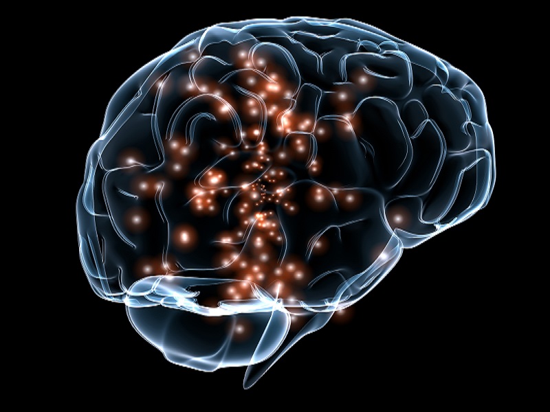
For some of us seasonal changes have a dramatic effect on our bodies. For others, it’s a subtle shift, if there’s one at all. This has to do with our circadian rhythms.

Our circadian rhythm is essentially an internal body clock. “[It] regulates our body with respect to sleeping, feeding and well-being,” Serani said. Circadian rhythms respond to sunlight. With less sun exposure in the fall and winter, many people experience a shift in their circadian rhythm, she said.

How can you tell if you’ve been affected? If you’re sluggish during the times of the day you used to feel energetic or you’re exhausted when you used to be well rested, the seasonal changes might be affecting you, she said.

To reset your clock, on the weekends, when possible, wake up without an alarm so your body gets adequate rest, Serani said. For some, melatonin supplements might improve sleep, she said.

Getting enough sunshine is key. Twenty minutes a day seems to be the magic number, Serani said. You can achieve that by going outside or soaking in the sunshine by a window, she said. Or you can buy a light box, which emits bright artificial light.

(Light therapy is actually very helpful for people with seasonal affective disorder, a form of clinical depression that occurs during the winter. This New York Times article has some good information.)

2. Keep up regular physical activity.

Depending on where you live, you might want to participate in winter activities, such as skiing, snowboarding, snowshoeing, ice-skating or hockey.

But if those aren’t appealing, Solomon said, “even taking a short walk each day or going to an indoor yoga class can help.” Workout DVDs are another option.

If you’re not sure what you like, try a variety of activities that sound like fun. Then pay attention to which activities boost your mood and energy levels.

3. Eat a variety of foods. 

“Make sure you’re eating a variety of foods, including as many fruits and vegetables as you can,” Solomon said. If fresh produce isn’t available, eat foods that are in season, she said.

Also, “Though the colder weather makes us crave sweets and starches, be mindful to keep protein in your diet as a balance,” Serani said. “Protein doesn’t spike your sugar levels, leaving you to feel more satisfied, less irritable and tired than simple carbohydrates and sugars.”

Keep in mind that this isn’t about restricting what you eat or feeling ashamed – or sinful – about eating sugar. (There’s nothing criminal about savoring your favorite desserts.) Rather, it’s about paying attention to how foods affect you, giving your body the nutrients it needs and enjoying what you eat.

4. Socialize.

As the temps take a nosedive, the last thing you might want to do is leave your house. But try. “Schedule regular contact with friends and family, even if it’s via Skype,” Solomon said. Still, make sure you’re also getting out, she stressed.

5. Pamper yourself.

When you think of treating yourself, what comes to mind? For instance, consider taking fragrant baths, drinking hot tea, reading books, lighting candles or cuddling with a loved one, said Serani, who tends to pamper herself more during the fall and wintertime. “These seasonal things raise dopamine, serotonin and oxytocin, feel-good hormones that improve mood,” she said.

Source: Psychentral.com


Syphilis’ (Sexually Transmitted Infection) origins still unknown

Researchers have said that despite trying to find the origins of the sexually transmitted infection syphilis they have come up with an empty hand.

According to the “Columbian” theory, Christopher Columbus’ crews brought the infection from America to Europe while returning home in 1492 and the first recorded epidemic of syphilis occurred, during the French invasion of the Italian city of Naples in 1495.

However, critic’s claim that the disease may have been present in Europe before Columbus’ return, and it just wasn’t distinguished from other conditions like leprosy until 1495, LiveScience reported.

Syphilis is capable of damaging heart, brain, eyes and bones, and can even be fatal if left untreated.

The first mention of the disease appears in the historical record in the 1496 writings of a man who went by the name Joseph Grnpeck.

However, Italian physician and poet Girolamo Fracastoro was the first man to use the word “syphilis” in 1530 in a Latin poem.

The study has been published in the Journal of Sexual Medicine.

Source: Yahoo News


Blame your genes for your aging brain

Senior author John Blangero, Ph.D., a Texas Biomed geneticist, said that identification of genes associated with brain aging should improve our understanding of the biological processes that govern normal age-related decline.

In large pedigrees including 1,129 people aged 18 to 83, the scientists documented profound aging effects from young adulthood to old age, on neurocognitive ability and brain white matter measures. White matter actively affects how the brain learns and functions.

Genetic material shared amongst biological relatives appears to predict the observed changes in brain function with age.

Blangero said that the use of large human pedigrees provides a powerful resource for measuring how genetic factors change with age.

By applying a sophisticated analysis, the scientists demonstrated a heritable basis for neurocognitive deterioration with age that could be attributed to genetic factors. Similarly, decreasing white matter integrity with age was influenced by genes.

The investigators further demonstrated that different sets of genes are responsible for these two biological aging processes

Source: Newstrackindia


Hair regeneration method uses patient’s own cells to grow new hair

Researchers at Columbia University Medical Center (CUMC) have devised a hair restoration method that can generate new human hair growth, rather than simply redistribute hair from one part of the scalp to another. The approach could significantly expand the use of hair transplantation to women with hair loss, who tend to have insufficient donor hair, as well as to men in early stages of baldness. The study was published today in the online edition of the Proceedings of the National Academy of Sciences (PNAS).

“About 90 percent of women with hair loss are not strong candidates for hair transplantation surgery because of insufficient donor hair,” said co-study leader Angela M. Christiano, PhD, the Richard and Mildred Rhodebeck Professor of Dermatology and professor of genetics & development. “This method offers the possibility of inducing large numbers of hair follicles or rejuvenating existing hair follicles, starting with cells grown from just a few hundred donor hairs. It could make hair transplantation available to individuals with a limited number of follicles, including those with female-pattern hair loss, scarring alopecia, and hair loss due to burns.”

According to Dr. Christiano, such patients gain little benefit from existing hair-loss medications, which tend to slow the rate of hair loss but usually do not stimulate robust new hair growth.

“Dermal papilla cells give rise to hair follicles, and the notion of cloning hair follicles using inductive dermal papilla cells has been around for 40 years or so,” said co-study leader Colin Jahoda, PhD, professor of stem cell sciences at Durham University, England, and co-director of North East England Stem Cell Institute, who is one of the early founders of the field.  “However, once the dermal papilla cells are put into conventional, two-dimensional tissue culture, they revert to basic skin cells and lose their ability to produce hair follicles.  So we were faced with a Catch-22: how to expand a sufficiently large number of cells for hair regeneration while retaining their inductive properties.”

The researchers found a clue to overcoming this barrier in their observations of rodent hair.  Rodent papillae can be easily harvested, expanded, and successfully transplanted back into rodent skin, a method pioneered by Dr. Jahoda several years ago. The main reason that rodent hair is readily transplantable, the researchers suspected, is that their dermal papillae (unlike human papillae) tend to spontaneously aggregate, or form clumps, in tissue culture. The team reasoned that these aggregations must create their own extracellular environment, which allows the papillae to interact and release signals that ultimately reprogram the recipient skin to grow new follicles.

“This suggested that if we cultured human papillae in such a way as to encourage them to aggregate the way rodent cells do spontaneously, it could create the conditions needed to induce hair growth in human skin,” said first author Claire A. Higgins, PhD, associate research scientist.

To test their hypothesis, the researchers harvested dermal papillae from seven human donors and cloned the cells in tissue culture; no additional growth factors were added to the cultures. After a few days, the cultured papillae were transplanted between the dermis and epidermis of human skin that had been grafted onto the backs of mice. In five of the seven tests, the transplants resulted in new hair growth that lasted at least six weeks. DNA analysis confirmed that the new hair follicles were human and genetically matched the donors.

“This approach has the potential to transform the medical treatment of hair loss,” said Dr. Christiano. “Current hair-loss medications tend to slow the loss of hair follicles or potentially stimulate the growth of existing hairs, but they do not create new hair follicles. Neither do conventional hair transplants, which relocate a set number of hairs from the back of the scalp to the front. Our method, in contrast, has the potential to actually grow new follicles using a patient’s own cells. This could greatly expand the utility of hair restoration surgery to women and to younger patients—now it is largely restricted to the treatment of male-pattern baldness in patients with stable disease.”

More work needs to be done before the method can be tested in humans, according to the researchers. “We need to establish the origins of the critical intrinsic properties of the newly induced hairs, such as their hair cycle kinetics, color, angle, positioning, and texture” said Dr. Jahoda. “We also need to establish the role of the host epidermal cells that the dermal papilla cells interact with, to make the new structures.”  The team is optimistic that clinical trials could begin in the near future. “We also think that this study is an important step toward the goal of creating a replacement skin that contains hair follicles for use with, for example, burn patients,” said Dr Jahoda.

The researchers used gene expression analyses to determine that the three-dimensional cultures restored 22 percent of the gene expression seen in normal hair follicles. “That’s less than we expected, but it was sufficient for inducing the growth of new hair follicles,” said Dr. Christiano.

In addition, using methods for the analysis of regulatory networks developed by the Califano lab in the Center for Computational Biology and Bioinformatics, Department of Systems Biology, the researchers identified a number of transcription factors (gene regulators) that have the potential to mimic the environmental signals that trigger papillae to induce new hair growth. This information could help researchers develop ways to restore the expression of more genes involved in hair growth and to increase the efficiency of the induction.

The paper is titled, “Microenvironmental reprogramming by three-dimensional culture enables dermal papilla cells to induce de novo human hair follicle growth.” The other contributors are James C. Chen and Jane E. Cerise, both at CUMC.

The study was supported by a Science of Human Appearance Career Development Award from the Dermatology Foundation and by grants from the Medical Research Council of the UK, the Empire State Development’s Division of Science, Technology and Innovation (NYSTAR), New York Stem Cell Science (NYSTEM), and the Biotechnology and Biological Sciences Research Council in the UK (BBSRC), as well as earlier support from the Steven and Michele Kirsch Foundation. Dr. Christiano is a member of the Columbia Stem Cell Initiative. The authors declare no financial or other conflicts of interests.

Source: Newsroom.columbia.edu


Cancer vaccine developed to boost lifespan of patients

Russian scientists have developed a vaccine for the treatment of cancer that can increase the patient’s lifespan more than two-fold, ITAR-TASS reported on Wednesday.

The vaccine, developed at the Institute of Clinical Immunology in the Siberian branch of the Russian Academy of Medical Sciences in Novosibirsk, has already successfully passed clinical tests, said institute’s director Vladimir Kozlov.

Currently, it is being administered to patients by injection at the third and fourth stages of cancer.

”We are deriving dendritic cells from the human body and loading them with tumor antigens,” Kozlov said.

“Dendritic cells process them, then we inject the cells into the patient and they start working in the body evoking a strong immune response. That is, they are actively fighting the tumor.”

The institute’s creation “is not a classical vaccine, which is the means to prevent the illness”, Kozlov said. “In fact, it is a cell therapy.”

The vaccine is counteracting several types of cancer – colorectal (bowel) cancer, breast cancer and prostate cancer – and the institute is ready to start its industrial production, Kozlov added.

However, the scientist added that other types of therapy were to be used as well, such as chemotherapy and radiation therapy.

Source: Khaleej Times


Apollo Hospitals launches web-based Personal Health Record

Hyderabad: Apollo Hospitals, in association with HealthHiway, on Tuesday launched Apollo Prism, a web-based personal health record (PHR) that would enable patients to receive, create and manage their health information such as lab test results, discharge summary, medical history, medications and other health interventions. The patients can then choose to share all or part of their health information with their loved ones and Apollo hospital.

Apollo Prism has been launched across all Apollo Hospitals locations. It has been tightly integrated with the hospital information system across locations, thus providing clinically validated information to patients, who come to Apollo Hospitals. Information is entirely patient controlled and completely confidential. Patients can also download the mobile application of Apollo Prism that lets them receive their lab test results on their phone. Apollo Prism is also an interface for diabetic patients, for managing their diabetes. Thus, beyond helping a patient to track and manage their health information, the PHR enables continuous, preventive and wellness support, according to a statement by Apollo Hospitals.

Sangita Reddy, executive director, Apollo Hospitals, said, “Apollo Prism is a significant step forward in our mission to further engage, involve and create a more delightful experience for our patients. When our patients have easy and secure access to their medical information, it reduces costs, improves outcomes and enables superior care.”

Madhu Aravind, CEO, HealthHiway, said, “Traditionally, care provided by a hospital was inward focused and physician centric. A PHR is a first step in involving and engaging the patient and makes the care delivery centered on the needs of the patient.”

Headquartered in Bangalore, HealthHiway is a cloud-based health information network that enables seamless exchange of health information – data sharing and transactional – between hospitals, clinics, diagnostics, insurers, doctors and patients.

Source: Indian Medical Times


Open-heart surgery gives new lease of life to 5-day-old baby

A five-day-old baby with G6PD deficiency – a disorder caused by an abnormal X-chromosome gene, successfully underwent a complex open-heart surgery recently.

According to medical literature, till date, only two paediatric cardiac cases were associated with the disorders have been successfully treated.

Dr Vijay Agarwal, chief paediatric cardiac surgeon, at Fortis Hospital, said, “The child had turned blue due to lack of oxygen supply in his body. His tests showed that he had transposition of great arteries that required immediate surgery if we want to save him.”

Luckily, Dr Agarwal said the hospital where the child was born had done a battery of tests as a part of their routine which revealed that he was suffering from G6PD.

“It was a crucial piece of information that helped us in taking the right measures. In G6PD patients, you have to avoid certain medications that may lead to heavy bleeding and death.
Therefore, it was the biggest challenge in front us,” said Dr Agarwal.

Dr Agarwal added that apart from the G6PD disorder the child also had a single coronary artery instead of two. “Usually in open-heart surgery we need to cool the child below 30 degree Celsius to protect his organs. In his case, it was not possible as that would have led to severe bleeding because of his disorder,” said Dr Agarwal.

Taking help of medical literature, the team of doctors took measures for all possible risk associated with the surgery. The surgery lasted for seven hours. “We avoided the general drugs used in anaesthesia. We maintained the cooling temperature at 33 degree Celsius. Luckily, everything fell in place and the operation was successful. The child had an unusual combination of problems. Moreover, the child weighed only 2.4kg and had jaundice at birth. He has still managed to survive and we are happy that he is now fit to be discharged,” said Dr Agarwal. The team of doctors will be presenting the case in international conferences because of its uniqueness.

Source: http://bit.ly/1doePCr