
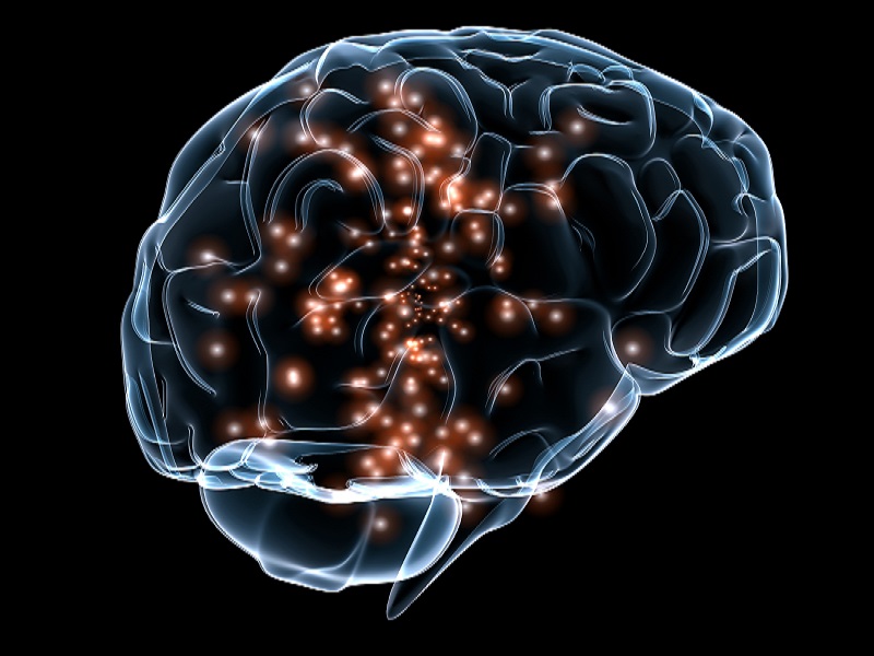
A recent study, published by researchers from the University of Kentucky, in the journal Free Radical Biology and Medicine, suggests that low levels of vitamin D may cause brain damage.
Vitamin D is a fat-soluble vitamin present in few natural foods, including fatty fish, cheese and egg yolks; a variety of foods, meanwhile, are artificially fortified with vitamin D, including milk, cereals and margarine. Vitamin D manufacture can also be achieved endogenously, when rays of light strike the skin. This photochemical process triggers the production of vitamin D3 (a.k.a. cholecalciferol) from its precursor, 7-dehydrocholesterol.
Vitamin D confers a number of benefits, ranging from promotion of calcium absorption in the gut, maintaining serum calcium and phosphate levels, as well as bone growth and remodeling. On top of this, vitamin D regulates a number of genes that are involved in cell division, differentiation and an essential form of programmed cellular death (apoptosis). It is thought that vitamin D serves a variety of roles in inflammatory processes and could even serve neuromuscular and immune functions.
The Rat Research Models
The latest scientific research suggests that the vitamin may serve a critical role in protecting the brain from free radical-induced damage. The researchers used a series of rat models to test the influence of differing concentrations of dietary vitamin D. A total of 27 male rats were divided into three separate groups; one group was fed a diet that contained low concentrations of vitamin D (100 IU/kg food), another was used as a control (1000 IU/kg food) and the final group received a diet enriched in the vitamin (10,000 IU/kg food).
The trial began as the rats hit middle-age and lasted for a period of four to five months. The research group measured the level of oxidative and nitrosative stress in a specific part of the rat brains, located in the posterior cortex.
Intriguingly, the group found an elevation in the level of a reactive nitrogen species, called 3-nitrotyrosine, in those rats that had received inadequate levels of vitamin D. Nitrotyrosine is considered a marker of cellular damage and inflammation and has been found to be elevated in a number of pathologies, including inflammatory diseases, lung disease, sepsis and atherosclerosis.
The researchers believe that the increase in nitrotyrosine is caused by disruption of a protein complex (NF-?B) that is recruited during cellular stress. In addition, after performing redox proteomics, a number of proteins in this region of the brain were found to be damaged in those rats that were provisioned low vitamin D diets.
When examining the real-world affect that this vitamin D deficiency had on the rats, the research team established that subjects provided with an abundance of the sunshine vitamin excelled in cognitive performance tests. Specifically, when investigating learning and memory capacity, rats given diets consisting of 100 IU/kg of food were found to lag behind the other two groups, significantly.
The Future
Allan Butterfield was the lead author of the latest study, who works as a professor in the UK Department of Chemistry and as the director of the Center of Membrane Sciences, faculty of Sanders-Brown Center on Aging. Also acting as the director of the Free Radical Biology in Cancer Core of the Markey Cancer Center, Butterfield briefly discussed his research endeavors and what they could mean for elderly populations.
“Given that vitamin D deficiency is especially widespread among the elderly, we investigated how during aging from middle-age to old-age how low vitamin D affected the oxidative the oxidative status of the brain… Adequate vitamin D serum levels are necessary to prevent free radical damage to the brain and subsequent deleterious consequences.”
This problem is exacerbated in developing countries, where food nutrition is problematic. Likewise, individuals that inhabit regions that receive little sunlight and those who work indoors for long periods are also prone to deficiency, as are elderly people who lead sedentary lifestyles.
In the past, prior scientific studies have implicated hypovitaminosis D in Alzheimer’s disease, with a number of researchers suggesting its use as a biomarker of disease progression. A recent study, published in the Journal of Alzheimer’s Disease, entitled Low serum vitamin D concentrations in Alzheimer’s disease: a systematic review and meta-analysis, found there to be low serum vitamin D levels in Alzheimer’s patients, relative to controls.
Meanwhile, a study produced by Lu’o’ng and Nguyen, which explored the beneficial role of vitamin D in Alzheimer’s patients, found that its absence could trigger mood problems and cognitive impairment. They also point to evidence that links vitamin D deficiency to a number of proteins that are adversely affected in Alzheimer’s disease pathology.
Aside from neurological disease, a number of studies have linked deficiency of the vitamin to the development of cancers and cardiovascular pathologies. Osteomalacia and rickets, witnessed in adults and children, respectively, are commonly documented complications of vitamin D deficiency, causing softening and bowing of bones.
In concluding, Butterfield recommends people consult their general practitioners to determine their vitamin D levels, eat food enriched in vitamin D and get a minimum of 10 to 15 minutes of sun exposure every day; he also suggests individuals prone to deficiency should ask their doctor for advice about taking vitamin D supplements.
Source: Guardian express


 With the city dwellers spending a greater time outside the home, either at work, or in traffic or in social networking sites, there is lesser interaction happening with one’s own relatives, said Dr Mahesh R Gowda, consultant physiatrist, Spandana health care.
 Our mouths are a delicate balance of good and bad bacteria. When we clean our teeth, the aim is to knock out cavity-causing bacteria, while allowing beneficial oral bacteria to thrive. Now, researchers have developed a sugar-free candy, which contains dead bacteria that bind to bad bacteria, potentially reducing cavities.
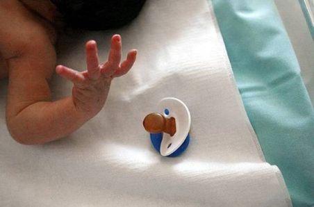 Researchers have claimed that babies dying from Sudden infant death syndrome (SIDS) have brain stem abnormalities regardless of whether they were exposed to risks like suffocation or co-sleeping.
 A new study by Indian origin researcher has revealed that the synthesis of the most active component of grape seed extract, B2G2, encourages the cell death known as apoptosis in prostate cancer cells while leaving healthy cells unharmed.


 A new research has showed that poor sleep quality is strongly associated with mood disturbance and lower quality of life among people with extreme obesity.