Coffee Cravings May Spring From Your DNA

Genes appear to influence how much caffeine you need

Anybody up for a steaming cup of Joe? Turns out your DNA may hold the answer.

New research suggests that your genes influence how much coffee you drink.

Researchers analyzed genetic data from more than 1,200 people in Italy, who were asked how much coffee they drank each day.

Those with a gene variant called PDSS2 drank one cup less a day on average than those without the variation, the investigators found.

Research involving more than 1,700 people in the Netherlands yielded similar findings, according to the study authors.

The findings suggest that PDSS2 reduces cells’ ability to break down caffeine. That means it stays in the body longer.

The upshot: People with the gene variant don’t need as much coffee to get the same caffeine hit as those without it, the researchers said.

“The results of our study add to existing research suggesting that our drive to drink coffee may be embedded in our genes,” said study author Nicola Pirastu. He is a chancellor’s fellow at the University of Edinburgh in Scotland.

“We need to do larger studies to confirm the discovery and also to clarify the biological link between PDSS2 and coffee consumption,” Pirastu added in a university news release.

By Robert Preidt

Source: https://medlineplus.gov/news/fullstory_160628.html


mHealth solutions: the future of health care

Through cloud computing, people can have seamless access to shared data, resources and common infrastructure.Over the network, organizations can offer services on demand and carry out tasks that meet changing needs and standards. Electronic applications make it possible to do all this, and more, in the health care setting.

Mobile health, or mHealth, incorporates cloud computing technology and devices such as tablets, mobile phones and personal digital assistants (PDAs) for a variety of purposes.But while it can make eHealth applications and medical information available anywhere at anytime, it must also be portable, secure and easy to use.

The range of applications and services supported by mHealth systems include:

  • Mobile telemedicine, used for remote consultations
  • Storing and sharing of patient data
  • Personalized monitoring of vitals, now enhanced through interconnectedness with wearable devices
  • Location-based medical services to ensure delivery of locally-relevant information
  • Emergency response and management
  • Pervasive access to health care information
  • But as mobile technology gathers pace, the possibilities may be limited only to our imagination

Advantages and challenges of mobile technology in health care

As governments and individuals experience ever-greater pressure to increase efficiency, mHealth solutions can offer numerous advantages.The mobility of an interconnected, wireless system means that it can be used anywhere, and specifically at the point of care.

Collaboration can reduce the risk of errors: there is less physical paperwork to get lost and a reduced risk of two doctors making different decisions.Point-of-care digital tools can help to safeguard patients and protect professionals against litigation through instant recording of data and potential for verification in real time and in the future.

mHealth can save time and money by enabling instant recording of information and a reduction in the duplication of tasks. It can enable virtual meetings, eliminating the need to move physically to a new location.Pooling of data and resources can lead to closer collaboration and stronger teams. Professional development becomes more feasible due to instant, online delivery of research, training materials and other updates.

The challenges of mHealth solutions include the practicalities of data storage and management, availability and maintenance of the network, as well as compatibility and interoperability.The biggest issue is perhaps security and privacy, raising questions about permission control, data anonymity and confidentiality, as well as the integrity of the infrastructure.The initial financial outlay and training and resistance to change within an organization may pose further challenges.

Source: medicalnews today


Middle-Age Heart Fitness Tied to Later Brain Health

Poorer heart health in middle age was tied to worse outcomes for the brain 20 years later, an observational study of Framingham Offspring participants has shown.
Poor cardiovascular (CV) fitness and greater diastolic blood pressure (BP) and heart rate (HR) response to exercise were associated with a smaller total cerebral brain volume (TCBV) almost 2 decades later (all P<0.05), Nicole L. Spartano, PhD, from the the Whitaker Cardiovascular Institute at Boston University, and colleagues reported online in Neurology.

The study also showed that each standard deviation in less fitness was equivalent to approximately 1 additional year of brain aging in those free of heart disease, they reported.

“Our investigation provides new evidence that lower CV fitness and elevated exercise BP and HR responses in early to midlife are associated with smaller brain volumes nearly 2 decades later, thereby linking fitness over the life course to brain health in later life,” wrote the investigators. “Promotion of midlife CV fitness may be an important step towards ensuring healthy brain aging in the population, especially in prehypertensive or hypertensive individuals.”

The study looked at men and women who were offspring (n=3,548) and spouses (n=1,576) of the original Framingham Heart Study cohort. Participants in the Framingham Offspring Study have had regular clinical examinations approximately every 4 years.
Between 1979 and 1983, at the baseline examination, Framingham Offspring participants with an average age of 40 years had an exercise treadmill test. Twenty years later (1998–2001), at the follow-up examination, participants — now an average age of 58 years old — had an abbreviated treadmill test as well as MRI brain scans.
Spartano and colleagues conducted two analyses. The primary analysis looked at 1,094 Framingham Offspring participants free from dementia and cardiovascular disease at baseline. More than half were female.

The secondary analysis included 1,583 Framingham Offspring participants who had cardiovascular disease or who were taking beta blockers after the baseline exam.
In the baseline exam, 89% of participants overall were able to achieve their target HR (85% of predicted HR maximum or VO2 max), with an estimated exercise capacity equivalent to 39 mL/kg/min.

Over the 19-year follow-up period, the prevalence of hypertension rose from 9% to 28%, and 60% of participants overall had either prehypertensive or hypertensive blood pressure, the study showed.

Previous studies have provided evidence of an association between exaggerated exercise BP and target organ damage or cardiovascular events, noted Spartano and colleagues.

“Individuals with exaggerated BP response to low levels of exercise may have vascular dysfunction that may not be discernible with examination of resting BP,” they wrote. “There is also growing evidence that ambulatory BP is more strongly associated with functional and structural brain impairments than resting BP measured in a clinical setting.”

They also pointed out that drug treatment of hypertension in older age has failed to show prevention of brain volume loss, but there is evidence that treatment of BP in midlife may prevent cognitive decline in later life.

While subtle structural brain changes may precede detectable cognitive impairment by up to a decade, the Coronary Artery Risk Development in Young Adults Study (CARDIA) study observed that physical fitness in early adulthood (mean age 25 years) was associated with cognition in later life (mean age 49 years).

“We are unable to account for the differences between our results and the results from the CARDIA study,” Spartano and colleagues wrote, adding that the effects of lower CV fitness may be more discernible in early adulthood.

“Our findings warrant confirmation in future investigations,” they said, noting that the cohort consisted mostly of white individuals of European descent, and that brain MRI measures were only available in later life.

“This study adds to the growing evidence that mid-life healthy lifestyle habits have an effect on brain aging decades later,” said Serge Gauthier, MD, of the McGill Centre for Studies in Aging and the Douglas Mental Health Research Institute in Montreal, who was not involved in the study.

“This does not prove that you can prevent dementia simply by doing physical exercise,” Gauthier told MedPage Today. “However, it justifies long-term randomized studies in populations with different levels of risk of cognitive decline with age, possibly combining different modalities such as cognitive training, diet, and social interaction.”

Source: medpagetoday


Texting at night affects teens’ sleep, academic performance

Can’t stop texting? If you’re a teenager, it may be to blame for falling grades and increased yawning in school, according to a new Rutgers study.

The study, published in the Journal of Child Neurology, is the first of its kind to link nighttime instant messaging habits of American teenagers to sleep health and school performance.

“We need to be aware that teenagers are using electronic devices excessively and have a unique physiology,” says study author Xue Ming, professor of neuroscience and neurology at Rutgers New Jersey Medical School. “They tend to go to sleep late and get up late. When we go against that natural rhythm, students become less efficient.”

The American Academy of Pediatrics reports that media use among children of all ages is increasing exponentially; studies have found that children ages 8 to 18 use electronic devices approximately seven-and-a-half hours daily.

Ming’s research is part of a small but growing body of evidence on the negative effects of electronics on sleep and school performance. But few studies, Ming says, have focused specifically on instant messaging.

“During the last few years I have noticed an increased use of smartphones by my patients with sleep problems,” Ming says. “I wanted to isolate how messaging alone – especially after the lights are out – contributes to sleep-related problems and academic performance.”

To conduct her study, Ming distributed surveys to three New Jersey high schools – a suburban and an urban public school and a private school – and evaluated the 1,537 responses contrasting grades, sexes, messaging duration and whether the texting occurred before or after lights out.

She found that students who turned off their devices or who messaged for less than 30 minutes after lights out performed significantly better in school than those who messaged for more than 30 minutes after lights out.

Students who texted longer in the dark also slept fewer hours and were sleepier during the day than those who stopped messaging when they went to bed. Texting before lights out did not affect academic performance, the study found.

Although females reported more messaging overall and more daytime sleepiness, they had better academic performance than males. “I attribute this to the fact that the girls texted primarily before turning off the light,” Ming says.

The effects of “blue light” emitted from smartphones and tablets are intensified when viewed in a dark room, Ming says. This short wavelength light can have a strong impact on daytime sleepiness symptoms since it can delay melatonin release, making it more difficult to fall asleep – even when seen through closed eyelids.

“When we turn the lights off, it should be to make a gradual transition from wakefulness to sleep,” Ming says. “If a person keeps getting text messages with alerts and light emission, that also can disrupt his circadian rhythm. Rapid Eye Movement sleep is the period during sleep most important to learning, memory consolidation and social adjustment in adolescents. When falling asleep is delayed but rising time is not, REM sleep will be cut short, which can affect learning and memory.”

Ming notes some benefits to early-evening media use, such as facilitating collaboration for school projects, providing resources for tutoring, increasing school readiness and possibly offering emotional support systems.

She suggests that educators recognize the sleep needs of teenagers and incorporate sleep education in their curriculum. “Sleep is not a luxury; it’s a biological necessity. Adolescents are not receiving the optimal amount of sleep; they should be getting 8-and-a-half hours a night,” says Ming. “Sleep deprivation is a strong argument in favor of later start times for high schools – like 9 a.m.”

Source: Sciencedaily


Simple injection to cure glaucoma

Glaucoma, an eye disease that damages the optic nerve and can result in vision loss and blindness, can now be treated with a simple injection, eye surgeons have said.

Simple injection to cure glaucoma

XenIn glaucoma, eye pressure plays a role in damaging the optic nerve.

A new technology developed by a US-based company relieves the pressure in the eye with a minimally invasive method, thus doing away with the need for scalpels and stitches, the experts noted.

It involves using a hypodermic needle to inject a tiny pliable drainage tube into the eye, allowing excess fluid to flow out, the Daily Mail reported.

The Xen Gel Stent developed by California-based AqueSys Inc is six mm long and about the width of a human hair. It is injected through a small self-sealing corneal incision using a simple, preloaded injector.

It was “quicker and safer” than current methods, Vik Sharma, a consultant eye surgeon at the Royal Free Hospital in London, was quoted as saying.

But the XEN Gel Stent is an investigational device, which means it has not been approved by the US Food and Drug Administration (FDA) yet.

The current approach in the management of patients with glaucoma begins with medications followed by laser surgery, or trabeculectomy, a treatment that frequently involves installing a small rigid tube in the eye to drain it.

This is attached to a metal plate embedded below the eye, which helps draw the fluid away. The eye must be cut to insert these relatively bulky items, and stitched up again, which leads to a high complication rate.

By comparison, the Xen Gel Stent is delivered directly via a hypodermic needle into the eye which has potential to lower complications rate compared to traditional methods.

source: India Medical Times


Reaction to coffee in your genes

Caffeine affects different people in different ways—and genetics could be the reason. Scientists have previously believed there is a genetic connection between individual responses to caffeine, but singling out the specific genetic variants has been a challenge. A recent study published in Molecular Psychiatry, however, provides new insight.

Researchers from the Harvard School of Public Health and Brigham and Women’s Hospital in Boston performed a meta-analysis on 120,000 regular coffee drinkers. Participants came from different ancestries–American, European and African ancestry.

Two gene variants were identified in connection to caffeine metabolism: POR and ABCG2. Two other gene variants near genes BDNF and SLC6A4 were associated with the “reward” effect of caffeine Also, the genes GCKR and MLXIPL, which play a role in glucose and lipid metabolism, were connected to the metabolic and neurological effects of caffeine for the first time.

The study is believed to be a major step forward in the research of coffee effects. It could help scientists identify subgroups of people most likely to benefit from increasing their coffee intake and those who would be better off if they cut back.

Source: health central


Nobel Prize in Medicine 2014 awarded for discovering brain’s ‘inner GPS’ system

The Nobel Assembly at Karolinska Institutet on Monday decided to award the 2014 Nobel Prize in Physiology or Medicine with one half to John O´Keefe and the other half jointly to May-Britt Moser and Edvard I Moser for their discoveries of cells that constitute a positioning system in the brain.

Nobel Prize in Medicine 2014 awarded for discovering brain’s ‘inner GPS’ system

ohn O’Keefe was born in 1939 in New York, US. He is currently affiliated with the University College, London, UK. May-Britt Moser was born in 1963 in Fosnavag, Norway. She is currently affiliated with the Centre for Neural Computation, Trondheim, Norway. Edvard I Moser was born in 1962 in Alesund, Norway. He is currently affiliated with the Kavli Institute for Systems Neuroscience, Trondheim, Norway.

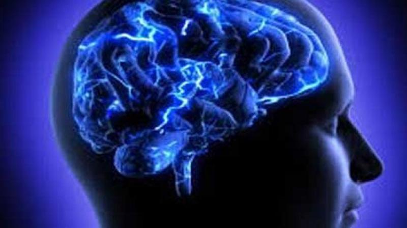
How do we know where we are? How can we find the way from one place to another? And how can we store this information in such a way that we can immediately find the way the next time we trace the same path? This year´s Nobel Laureates have discovered a positioning system, an “inner GPS” in the brain that makes it possible to orient ourselves in space, demonstrating a cellular basis for higher cognitive function.

In 1971, John O´Keefe discovered the first component of this positioning system. He found that a type of nerve cell in an area of the brain called the hippocampus that was always activated when a rat was at a certain place in a room. Other nerve cells were activated when the rat was at other places. O´Keefe concluded that these “place cells” formed a map of the room.

More than three decades later, in 2005, May-Britt and Edvard Moser discovered another key component of the brain’s positioning system. They identified another type of nerve cell, which they called “grid cells”, that generate a coordinate system and allow for precise positioning and pathfinding. Their subsequent research showed how place and grid cells make it possible to determine position and to navigate.

The discoveries of John O´Keefe, May-Britt Moser and Edvard Moser have solved a problem that has occupied philosophers and scientists for centuries – how does the brain create a map of the space surrounding us and how can we navigate our way through a complex environment?

How do we experience our environment?

The sense of place and the ability to navigate are fundamental to our existence. The sense of place gives a perception of position in the environment. During navigation, it is interlinked with a sense of distance that is based on motion and knowledge of previous positions.

Questions about place and navigation have engaged philosophers and scientists for a long time. More than 200 years ago, the German philosopher Immanuel Kant argued that some mental abilities exist as a priori knowledge, independent of experience. He considered the concept of space as an inbuilt principle of the mind, one through which the world is and must be perceived. With the advent of behavioural psychology in the mid-20th century, these questions could be addressed experimentally. When Edward Tolman examined rats moving through labyrinths, he found that they could learn how to navigate, and proposed that a “cognitive map” formed in the brain allowed them to find their way. But questions still lingered – how would such a map be represented in the brain?

John O´Keefe and the place in space

John O´Keefe was fascinated by the problem of how the brain controls behaviour and decided, in the late 1960s, to attack this question with neurophysiological methods. When recording signals from individual nerve cells in a part of the brain called the hippocampus, in rats moving freely in a room, O’Keefe discovered that certain nerve cells were activated when the animal assumed a particular place in the environment (Figure 1). He could demonstrate that these “place cells” were not merely registering visual input, but were building up an inner map of the environment. O’Keefe concluded that the hippocampus generates numerous maps, represented by the collective activity of place cells that are activated in different environments. Therefore, the memory of an environment can be stored as a specific combination of place cell activities in the hippocampus.

May-Britt and Edvard Moser find the coordinates

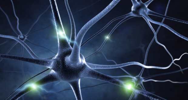
May-Britt and Edvard Moser were mapping the connections to the hippocampus in rats moving in a room when they discovered an astonishing pattern of activity in a nearby part of the brain called the entorhinal cortex. Here, certain cells were activated when the rat passed multiple locations arranged in a hexagonal grid (Figure 2). Each of these cells was activated in a unique spatial pattern and collectively these “grid cells” constitute a coordinate system that allows for spatial navigation. Together with other cells of the entorhinal cortex that recognize the direction of the head and the border of the room, they form circuits with the place cells in the hippocampus. This circuitry constitutes a comprehensive positioning system, an inner GPS, in the brain (Figure 3).

A place for maps in the human brain

Recent investigations with brain imaging techniques, as well as studies of patients undergoing neurosurgery, have provided evidence that place and grid cells exist also in humans. In patients with Alzheimer´s disease, the hippocampus and entorhinal cortex are frequently affected at an early stage, and these individuals often lose their way and cannot recognize the environment. Knowledge about the brain´s positioning system may, therefore, help us understand the mechanism underpinning the devastating spatial memory loss that affects people with this disease.

The discovery of the brain’s positioning system represents a paradigm shift in our understanding of how ensembles of specialized cells work together to execute higher cognitive functions. It has opened new avenues for understanding other cognitive processes, such as memory, thinking and planning.

Source: india medical times


New type of brain cell discovered

Researchers have discovered a strange new type of brain cell that sends signals by bypassing the cell body altogether. Neurons come in different shapes and sizes but the basic blueprint consists of a cell body, from which protrudes spindly appendages called dendrites and axons.

New type of brain cell discovered

Dendrites are branchlike structures that receive signals from other nerve cells and deliver them to the cell body. The neuron then processes the signals and zaps along information to the next cell via a long projection called the axon.

The newly discovered cells, however, have a different, and until now, unknown process. In these cells, the signals skip the cell body altogether, instead travelling along an axon that projects directly from one of the dendrites.

“We found that in more than half of the cells, the axon does not emerge from the cell body, but arises from a lower dendrite,” said study researcher Christian Thome, a neuroscientist at Heidelberg University and the Bernstein Center Heidelberg-Mannheim. The new cells were discovered in the hippocampus of a mouse. Humans have the same general brain structure and types of hippocampus cells as mice. The hippocampus is home to extensively branched neurons called pyramidal cells because of their triangular cell bodies, ‘Live Science’ reported.

Source: times of india


Research reveals how Ebola virus blocks immune system

The Ebola virus, in the midst of its biggest outbreak on record, is a master at evading the body’s immune system. But researchers at Washington University School of Medicine in St. Louis and elsewhere have learned one way the virus dodges the body’s antiviral defenses, providing important insight that could lead to new therapies.

Research reveals how Ebola virus blocks immune system

The virus has infected about 1,800 people since March in four West African nations and killed more than half of them, according to the World Health Organization.

The researchers developed a detailed map of how an Ebola protein, VP24, binds to a host protein that takes signaling molecules in and out of the cell nucleus. Their map revealed that the viral protein takes away the host protein’s ability to carry an important immune signal into the nucleus.

This signal helps activate the immune system’s antiviral defenses, and blocking it is believed to contribute significantly to the virus’s deadliness.

“We’ve known for a long time that infection with Ebola obstructs an important arm in our immune system that is activated by molecules called interferons,” said senior author Gaya Amarasinghe, PhD, assistant professor of pathology and immunology at the School of Medicine. “Now that our map of the combined structure of these two proteins has revealed one critical way Ebola does this, the information it provides will guide the development of new treatments.”

The results appear Aug. 13 in Cell Host & Microbe.

A National Institutes of Health (NIH) grant of up to $15 million, awarded March 1, is helping Amarasinghe and other researchers look for drugs to block VP24 and another Ebola protein, VP35. The group includes researchers at the Icahn School of Medicine at Mount Sinai, Washington University, the University of Texas Southwestern Medical Center, Howard University and Microbiotix Inc., a Massachussetts biopharmaceutical company.

Co-author Christopher Basler, PhD, professor of microbiology at Mount Sinai and the principal investigator of the consortium, was the first to show that VP24 and VP35 were important to the virus’s ability to keep the immune system at bay.

In an earlier paper, Amarasinghe, Basler and others revealed that VP35 blocks production of interferon, one of the main regulators of the innate immune system. This branch of the immune system specializes in fighting viruses.

“Interferon is critical to our ability to defend ourselves against viruses,” Basler said. “It makes a variety of responses to viral infection possible, including the self-destruction of infected cells and the blockage of supplies necessary for viral reproduction.”

In the new study, Amarasinghe and Daisy Leung, PhD, assistant professor of pathology and immunology at Washington University, showed that VP24 tightly binds to a nuclear transporter, a protein that takes molecules into and out of the cell nucleus. Among the molecules these transporters take into the nucleus is STAT1, an important component of the interferon signaling pathway.

“Normally STAT1 is transported into the nucleus and activates the genes for hundreds of proteins involved in antiviral responses,” Leung said. “But when VP24 is attached to some of these transporters, STAT1 can’t get into the nucleus.”

The researchers found that VP24’s action specifically prevents STAT1 transport. Other proteins that travel in and out of the cell nucleus and are important to viral replication likely are unaffected. The scientists already have initiated efforts to look for small molecules that block VP35 and now are applying those same approaches to VP24.

Source: washington university


New way to diagnose malaria by detecting parasite’s waste in infected blood cells

Over the past several decades, malaria diagnosis has changed very little. After taking a blood sample from a patient, a technician smears the blood across a glass slide, stains it with a special dye, and looks under a microscope for the Plasmodium parasite, which causes the disease. This approach gives an accurate count of how many parasites are in the blood — an important measure of disease severity — but is not ideal because there is potential for human error.

New way to diagnose malaria by detecting parasite's waste in infected blood cells

A research team from the Singapore-MIT Alliance for Research and Technology (SMART) has now come up with a possible alternative. The researchers have devised a way to use magnetic resonance relaxometry (MRR), a close cousin of magnetic resonance imaging (MRI), to detect a parasitic waste product in the blood of infected patients. This technique could offer a more reliable way to detect malaria, says Jongyoon Han, a professor of electrical engineering and biological engineering at MIT.

“There is real potential to make this into a field-deployable system, especially since you don’t need any kind of labels or dye. It’s based on a naturally occurring biomarker that does not require any biochemical processing of samples” says Han, one of the senior authors of a paper describing the technique in the Aug. 31 issue of Nature Medicine.
Peter Rainer Preiser of SMART and Nanyang Technical University in Singapore is also a senior author. The paper’s lead author is Weng Kung Peng, a research scientist at SMART.

Hunting malaria with magnets
With the traditional blood-smear technique, a technician stains the blood with a reagent that dyes cell nuclei. Red blood cells don’t have nuclei, so any that show up are presumed to belong to parasite cells. However, the technology and expertise needed to identify the parasite are not always available in some of the regions most affected by malaria, and technicians don’t always agree in their interpretations of the smears, Han says.

“There’s a lot of human-to-human variation regarding what counts as infected red blood cells versus some dust particles stuck on the plate. It really takes a lot of practice,” he says.

The new SMART system detects a parasitic waste product called hemozoin. When the parasites infect red blood cells, they feed on the nutrient-rich hemoglobin carried by the cells. As hemoglobin breaks down, it releases iron, which can be toxic, so the parasite converts the iron into hemozoin — a weakly paramagnetic crystallite.

Those crystals interfere with the normal magnetic spins of hydrogen atoms. When exposed to a powerful magnetic field, hydrogen atoms align their spins in the same direction. When a second, smaller field perturbs the atoms, they should all change their spins in synchrony — but if another magnetic particle, such as hemozoin, is present, this synchrony is disrupted through a process called relaxation. The more magnetic particles are present, the more quickly the synchrony is disrupted.

“What we are trying to really measure is how the hydrogen’s nuclear magnetic resonance is affected by the proximity of other magnetic particles,” Han says. For this study, the researchers used a 0.5-tesla magnet, much less expensive and powerful than the 2- or 3-tesla magnets typically required for MRI diagnostic imaging, which can cost up to $2 million. The current device prototype is small enough to sit on a table or lab bench, but the team is also working on a portable version that is about the size of a small electronic tablet.

After taking a blood sample and spinning it down to concentrate the red blood cells, the sample analysis takes less than a minute. Only about 10 microliters of blood is required, which can be obtained with a finger prick, making the procedure minimally invasive and much easier for health care workers than drawing blood intravenously.

“This system can be built at a very low cost, relative to the million-dollar MRI machines used in a hospital,” Peng says. “Furthermore, since this technique does not rely on expensive labeling with chemical reagents, we are able to get each diagnostic test done at a cost of less than 10 cents.”

Tracking infection
Hemozoin crystals are produced in all four stages of malaria infection, including the earliest stages, and are generated by all known species of the Plasmodium parasite. Also, the amount of hemozoin can reveal how severe the infection is, or whether it is responding to treatment. “There are a lot of scenarios where you want to see the number, rather than a yes or no answer,” Han says.

In this paper, the researchers showed that they could detect Plasmodium falciparum, the most dangerous form of the parasite, in blood cells grown in the lab. They also detected the parasite in red blood cells from mice infected with Plasmodium berghei.

The researchers are launching a company to make this technology available at an affordable price. The team is also running field tests in Southeast Asia and is exploring powering the device on solar energy, an important consideration for poor rural areas.

Source: science daily