Simple injection to cure glaucoma

Glaucoma, an eye disease that damages the optic nerve and can result in vision loss and blindness, can now be treated with a simple injection, eye surgeons have said.

Simple injection to cure glaucoma

XenIn glaucoma, eye pressure plays a role in damaging the optic nerve.

A new technology developed by a US-based company relieves the pressure in the eye with a minimally invasive method, thus doing away with the need for scalpels and stitches, the experts noted.

It involves using a hypodermic needle to inject a tiny pliable drainage tube into the eye, allowing excess fluid to flow out, the Daily Mail reported.

The Xen Gel Stent developed by California-based AqueSys Inc is six mm long and about the width of a human hair. It is injected through a small self-sealing corneal incision using a simple, preloaded injector.

It was “quicker and safer” than current methods, Vik Sharma, a consultant eye surgeon at the Royal Free Hospital in London, was quoted as saying.

But the XEN Gel Stent is an investigational device, which means it has not been approved by the US Food and Drug Administration (FDA) yet.

The current approach in the management of patients with glaucoma begins with medications followed by laser surgery, or trabeculectomy, a treatment that frequently involves installing a small rigid tube in the eye to drain it.

This is attached to a metal plate embedded below the eye, which helps draw the fluid away. The eye must be cut to insert these relatively bulky items, and stitched up again, which leads to a high complication rate.

By comparison, the Xen Gel Stent is delivered directly via a hypodermic needle into the eye which has potential to lower complications rate compared to traditional methods.

source: India Medical Times


New layer of cornea discovered by Indian doctor Harminder Dua

A new layer in the human cornea was discovered by the Indian doctor Harminder Dua at The University of Nottingham in 2014. It plays a vital role in the structure of the tissue that controls the flow of fluid from the eye.

The research was published in a paper in the British Journal of Ophthalmology.
The new finding could shed new light on glaucoma. The new layer named as Dua’s Layer is just 15 microns thick but incredibly tough. Comprised of thin plates of collagen, it sits at the back of the cornea between the corneal stroma and Descemet’s membrane.

It makes an important contribution to the sieve-like meshwork, the trabecular meshwork (TM), in the periphery of the cornea.

The TM is a wedge-shaped band of tissue that extends along the circumference of the angle of the anterior chamber of the eye.

It is made of beams of collagen wrapped in a basement membrane to which trabecular cells and endothelial cells attach. The beams branch out randomly to form a ‘meshwork’.
Scientists had previously believed the cornea to be comprised of five layers – from front to back— the corneal epithelium, Bowman’s layer, the corneal stroma, Descemet’s membrane and the corneal endothelium.

Pressure within the eye is maintained by the balance of aqueous fluid production by eye tissue called the ciliary body and drainage principally through the TM to the canal of Schlemm, a circular channel in the angle of the eye.

Defective drainage through the TM is an important cause of glaucoma, a condition that leads to raised pressure in the eye that can permanently affect sight. Around 1 to 2% of the world’s population yearly have chronic glaucoma and globally around 45 million people have open angle glaucoma which can permanently damage the optic nerve – 10% of whom are blind.

It is hoped the discovery will offer new clues on why the drainage system malfunctions in the eyes of some people, leading to high pressure.
Glaucoma is a devastating disease caused by defective drainage of fluid from the eye. Glaucoma is the second largest leading cause of blindness of world.

Source: Zee News


No More Eye Drops? New Contact Lens Delivers Glaucoma Meds

Like a miniature donut stuffed inside a tiny pita pocket, a common glaucoma medicine is sandwiched inside this specially designed contact lens. In laboratory experiments, the lens, which can also correct vision, releases the eyesight-saving medication at a steady rate for up to a month. Its construction offers numerous potential clinical advantages over the standard glaucoma treatment and may have additional applications, such as delivering anti-inflammatory drugs or antibiotics to the eye.

Glaucoma is a group of conditions that can result in irreversible blindness. This vision loss can be reduced if glaucoma is found and treated early, most commonly with eye drops to lower pressure within the eye. But using eye drops regularly can be a challenge. And while the drops can minimize further vision loss, they don’t repair vision that’s already lost.

People using traditional eye drops for glaucoma “aren’t getting any symptomatic relief, and they’re not seeing better, so there’s not a lot of motivation to be compliant with the medication,” said Joseph Ciolino, an ophthalmologist who, along with his mentor Daniel Kohane, developed the new contact lens at Harvard Medical School.

Source: Daily me


Electrical burn causes star-shaped cataracts in patient’s eyes

cataract

A 42-year-old electrician in California developed star-shaped cataracts in his eyes after a serious work-related accident caused electricity to run through his body, according to a new report of the case.

The man’s left shoulder came into contact with 14,000 volts of electricity, and an electric current passed through his entire body, including the optic nerve — the nerve that connects the back of the eye to the brain.

“The optic nerve is similar to any wire that conducts electricity,” said Dr. Bobby Korn, an associate professor of clinical ophthalmology at the University of California, San Diego, who treated the patient. “In this case, the extreme current and voltage that passed through this important natural wire caused damage to the optic nerve itself,” Korn said.

Four weeks after the accident, Korn evaluated the patient, who was experiencing vision problems. An examination showed the man had “striking cataracts in both of his eyes,” that were star-shaped, Korn said. A cataract is a clouding of the lens in the eye.

The reason cataracts sometimes take on a star shape is not fully understood, Korn said. In animal studies, damage to the eye’s lens from electricity first appears as small bubbles called vacuoles on the outside of the lens. These bubbles then coalesce to form a star-shaped cataract, Korn said.

Four months after the accident, the man had surgery to remove the cataracts and implant a new lens, and his vision improved slightly after the operation, Korn said. But the damage to his optic nerve still limited the man’s sight, Korn said.

Korn explained that the eye is like a camera: If the lens is damaged, it can be replaced with a new one, but if the “film” — in this case, the optic nerve and retina — is damaged, “then you’ll never get a good picture,” Korn said.

Now, 10 years later, the man still has poor vision in both of his eyes, Korn said. But he is able to commute on public transportation and take classes at a community college using assistance, Korn said.

The man’s case is reported in the Jan. 23 issue of the New England Journal of Medicine.

Source: NBC news


Sleeping on one side may worsen glaucoma: study

In a new study from South Korea, people with worsening glaucoma on just one side were also more likely to sleep with the affected eye facing downward.

The researchers say that position raises the eye’s internal pressure and probably hastens deterioration of the eye.

In glaucoma, the optic nerve is often damaged by increased intraocular pressure. The damage causes tunnel vision and eventual blindness.

According to the World Health Organization, glaucoma is the second leading cause of blindness around the world, after cataracts.

“There is prior data from the early nineties, suggesting that in patients with glaucoma who sleep on their sides, the eye in the dependent position tends to have greater damage of the optic nerve,” Dr. Jeffrey Schultz told Reuters Health in an email.

Schultz directs Glaucoma Service at the Montefiore Medical Center in New York and is an associate professor at the Albert Einstein College of Medicine.

He was not involved in the study, but said it is “important in letting us know that there is potential for behavior changes in lessening the risk of blindness from glaucoma.”

The study was led by Dr. Kyoung Nam Kim, a researcher in the Department of Ophthalmology at the Chungnam National University hospital in Daejeon.

Treatments to decrease pressure in a patient’s eyeballs can slow progression of glaucoma in some cases. But other patients continue to progress even when intraocular pressure appears to be under control, Kim’s team writes in the American Journal of Ophthalmology.

Since lying down raises the pressure in the eyeball, and sleeping on one side consistently more than the other could be problematic for the eye on that side, the researchers decided to investigate whether a side- sleeping position might be part of the problem.

Kim and colleagues examined the sleeping habits of 430 glaucoma patients who had a visual field loss that was worse in one eye.

They found 132 of the patients preferred to sleep on one side. Of these patients, 67 percent usually slept with the worse eye downward.

They also compared the sleeping habits of patients who had glaucoma with elevated intraocular pressure (high-tension glaucoma) with those with normal pressure (normal-tension glaucoma).

Approximately 66 percent of the patients with normal-tension glaucoma preferred to sleep with the worse eye downward and 71 percent of the patients with high-tension glaucoma slept that way.

The results don’t prove that sleeping position accounts for worsening glaucoma on one side.

But they at least verify a link “between the preferred sleeping position and asymmetric visual field loss between eyes,” the authors write.

“Unfortunately, it is very difficult to control your body position during sleep,” Schultz said.

“Certainly, if one has severe damage in one eye it would seem to make sense to attempt to avoid sleeping on your side with that eye down,” he said.

It may help to sleep on the side with less eye damage – or on your back. But Schultz warns that sleeping on your back may not be the answer for people who are predisposed to sleep apnea, which is another risk factor for worsening glaucoma.

At this point there is no way to improve visual field loss in patients with glaucoma once it occurs, he said.

“The best thing that patients can do to lessen the risk of worsening, is to be compliant with the medical regime and to follow up as directed by the patient’s physician,” Schultz said.

Source: Reuters


Gene therapy reverses blinding eye disease

An experimental therapy for a blinding eye disease showed early – and surprising – promise when it improved the vision of patients in an early trial that was only supposed to test its safety, doctors reported Wednesday.

The experimental gene therapy not only stopped the steady degeneration of the patients’ vision, but appears to have reversed some of the damage. And the effects have lasted two years in one case, British researchers report in the Lancet medical journal.

Wayne Thompson of Staffordshire in Britain saw the stars for the first time in years after being treated in April.

“One night in the summer, my wife called me outside as it was a particularly starry evening. As I looked up, I was amazed that I was able to see a few stars,” Thompson, 43, said in a statement.

“I hadn’t seen stars for a long, long time,” he added.

“It is still too early to know if the gene therapy treatment will last indefinitely, but we can say that the vision improvements have been maintained for as long as we have been following up the patients, which is two years in one case,” says Dr. Robert MacLaren of the Nuffield Laboratory of Ophthalmology at the University of Oxford, who leads the research team.

“In truth, we did not expect to see such dramatic improvements in visual acuity and so we contacted both patients’ home opticians to get current and historical data on their vision in former years, long before the gene therapy trial started. These readings confirmed exactly what we had seen,” he added in a statement.

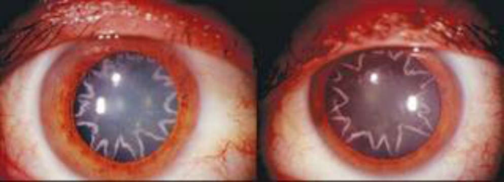
The men taking part in the trial all have choroideremia, a genetic disease that causes vision to start breaking down when patients are still children. The defective gene, called CHM, is on the x chromosome, so it almost exclusively affects males – females have an extra x chromosome that usually makes up for any lost function. It causes about 4 percent of all cases of blindness, according to the National Institutes of Health.

The condition breaks down various parts of the retina, the reflective tissue at the back of the eye that collects light and images. The gene therapy approach uses a virus to carry a corrective gene directly into the retina.

Gene therapy itself has a mixed record – it’s harder than scientists thought to safely deliver new genes into the body. And there is no guarantee that the patient’s cells will accept the new gene and use it.

But the eye is a good place to try, in part because doctors can see the effects in real time, and also because one eye can be treated and the other eye used to compare results.

“I think that this is a very important study,” said Dr. Richard Weleber, professor of ophthalmology who is leading a team at Oregon Health and Science University trying gene therapy to treat Usher Syndrome.

MacLaren’s trial was meant to be a stage 1 safety trial, designed simply to show that the treatment did no harm. But the six patients in the trial said they noticed improvements quickly.

“This has huge implications for anyone with a genetic retinal disease such as age-related macular degeneration or retinitis pigmentosa, because it has for the first time shown that gene therapy can be applied safely before the onset of vision loss,” MacLaren said.

He says nine patients have now had one eye treated with the gene therapy in operations at the Oxford Eye Hospital. The patients had varying degrees of vision loss, but MacLaren thinks the best course will be to treat people when they are young, before much damage has been done.

“If we were able to treat people early, get them in their teens or late childhood, we’d be getting the virus in before their vision is lost,” he said. “If the treatment works, we would be able to prevent them from going blind.”

Weleber, who says his own gene therapy trial is moving along but who could not release details, says MaCLaren’s surgical technique may have helped the therapy work more successfully. MacLaren’s team lifted an area called the macula to deliver the engineered viruses directly into the tissue that needed correcting.

Last year, researchers reported success against a different genetic disease causing blindness , one called Leber congenital amaurosis. But even though the patients’ vision improved, the eye continued to deteriorate.

“Each type (of genetic disease) needs to be tested separately,” Weleber said.

Gene therapy also works to some degree against blood cancers and immune diseases.

Last year, doctors reported they had successfully treated the first children in the U.S. for severe combined immune deficiency or SCID, sometimes known as “bubble boy” disease. Like choroideremia, SCID is caused by a mutated recessive gene, meaning children must inherit a defective copy from each parent to show symptoms.

Gene therapy has helped 26 of 59 two patients with a form of cancer called chronic lymphocytic leukemia go into remission after a type of gene therapy treatment that programmed immune system cells to hunt down and kill the leukemia cells. And last year doctors reported 24 people with acute lymphocytic leukemia got at least temporary remission after gene therapy.

Source: nbc news