Indian-Origin Scientist Paves Way For Better Epilepsy Treatments

epilepsy

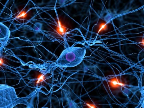
University of Toronto biologists, including one of Indian origin, have discovered proteins to retune imbalances of neurological disorders like autism, epilepsy and various others like schizophrenia and spectrum disorder.

According to Professor Melanie Woodin, the lead investigator of the study, there is a process known as synapses via which neurons in the brain correspond with other neurons, causing neurons either to excite or inhibit other neurons. He further added that any disproportion among the levels of excitation or inhibition may lead to improper brain function.

A crucial complex of protein has been identified that can regulate the proper correspondence of neurons at cellular level. The major proteins are KCC2 which is essential for inhibitory impulse, whereas the receptor for excitatory transmitter glutamate is GluK2, and Neto 2 protein interface with the other two proteins. All three proteins required for synaptic communication is brought together by this complex.

Vivek Madhavan, lead author of the study, along with other researchers conducted experiments on mice brain and found out that all the three proteins directly interact and control each others’ function.

BLUE NATIVE PAGE proved to be the most successful technique of applying a sensitive gel system for determining native protein complexes in neurons.

As such there is no treatment for epilepsy, and the treatments which are available can only curb its effects. Thus the main focus should be on its prevention.

Source: indian nerve


Useful safety tips for epilepsy patients

Safety in the home starts with your kitchen and your bathrooms. These are the places where, during and after any seizure, you can become confused and risk injury. Take these seizure precautions to decrease the chance of accidents.

Safeguard your kitchen

  • Use oven mitts and cook only on rear burners
  • If possible, use an electric stove, so there is no open flame
  • Cooking in a microwave is the safest option
  • Ask your plumber to install a heat-control device in your faucet so the water doesn’t become too hot
  • Carpet the kitchen floor. This can provide cushioning if you fall
  • Use plastic containers rather than glass when possible

Safeguard your bathroom

  • Install a device in your tub and showerhead that controls temperature. This keeps you from burning yourself if a seizure occurs
  • Carpet the floor—it’s softer and less slippery than tile
  • Do not put a lock on the bathroom door. If you have one, never use it. Someone should always be able to get in if you need help
  • Learn to bathe with only a few inches of water in the tub, or use a handheld showerhead
  • Planning ahead for safety outside the home

Driving.

For many people with epilepsy, the risk of seizures restricts their independence, in particular the ability to drive. The Epilepsy Foundation offers a state-by-state database of driving restrictions and regulations on its website. Find out more about driving and epilepsy.
Participating in activities.

You can play sports with epilepsy, but it’s a good idea to have someone with you who knows how to manage a seizure. Wearing head protection is also recommended when you participate in a contact sport that might cause you to fall or hit your head.

Here are some tips for picking the right physical activities when you are living with epilepsy:

  • If seizures usually occur at a certain time, plan activities when seizures are less likely to happen
  • Avoid extreme heat when exercising and keep hydrated with plenty of water to reduce seizure risks
  • Check with your neurologist before starting any new exercise program

Some activities may be restricted if you have uncontrolled seizures, including:

  • Swimming alone
  • Climbing to unsafe heights
  • Riding a bike in traffic

Source: VIMPAT


Famous Amnesia Patient’s Brain Cut into 2,401 Slices

Image of the frozen brain at the level of the temporal lobes during the cutting procedure.

A new examination of the brain of Patient H.M. — the man who became an iconic case in neuroscience when he developed a peculiar form of amnesia after parts of his brain were removed during surgery in 1953 — shows that his surgeon removed less of his brain than thought.

At age 27, H.M., whose real name was Henry Molaison, underwent an experimental surgical treatment for his debilitating epilepsy. His surgeon removed the medial temporal lobe, including a structure called the hippocampus.

Thereafter, H.M. was unable to form new memories. His case brought about the idea that the hippocampus may have a crucial role in retaining learned facts, replacing the notion that memories are scattered throughout the brain. H.M. became the focus of more than 50 years of memory research, working closely with the researchers who had to introduce themselves every time they met.

“Much of what we know about human memory, it has one way or another to do with H.M.,” said study researcher Jacopo Annese, director of The Brain Observatory in San Diego.

After H.M.’s death in 2008, Annese and his colleagues cut the patient’s frozen brain into 2,401 slices, each 0.7-millimeters thick. They took a picture of every slice, and created a high-resolution, 3D model of his brain.

In the new study detailed online today (Jan. 28) in the journal Nature Communications, they report that a significant portion of the hippocampus, which was thought to have been removed in surgery, was actually intact.

What happened to H.M.?

Research on H.M. showed that there are in fact different kinds of memory. He was unable to learn new facts, remember the events happening around him or learn people’s names, but he was able to recall events from his childhood. He also could learn skills, for example, he could get better at a new motor task with practice.

“Over 50 years of studies, the picture [of memory] was a little bit complicated,” because H.M. had some types of memory but not others, Annese said.

The only way to start teasing out H.M.’s memory impairment in light of the anatomy of the brain was to know what exactly had happened during the surgery

Until the 1990s, the researchers had only sketches drawn by the surgeon, Dr. William Scoville, to refer to. But after the advent of neuroimaging, researchers scanned H.M.’s brain in 1992 and found that a portion of the hippocampus had been spared.

In the new study, Annese and his colleagues measured the exact length of H.M.’s hippocampus, and found the spared portion was even larger than what brain scans had shown.

The posterior part of the hippocampus deals with memory, and the brain slices show this part wasn’t removed, and in fact, was undamaged at the cellular level, the researchers said.

“The most beautiful finding I think was the fact that we realized … that Scoville missed the posterior hippocampus,” Annese said.

The memory impairment

The new findings shed light on what happened to H.M., but likely won’t revolutionize what researchers know about memory, and are in fact in line with modern views of hippocampal function

Almost all connections from the hippocampus to the cortex go through a part of the temporal lobe called the entorhinal cortex, which Annese found had been removed from H.M.’s brain. As this region connects the hippocampus to other brain regions, the surgery may have nearly isolated the hippocampus from the rest of the brain.

This may mean that H.M.’s amnesia had more to do with the entorhinal cortex being removed, than with the parts of the hippocampus being removed, Annese said, although more study is needed to know for sure.

The new study presents “an extremely detailed post-mortem investigation of the remaining anatomy of [H.M.’s] brain,” said Neil Burgess, a memory researcher at University College London, who wasn’t involved in the new analysis. “These extra details will no doubt continue to fuel the debate as to which bits of the medial temporal lobe are responsible for which aspects of memory.”

Source: live science